They are ubiquitously produced—usually each cell type generates one or two dominant prostaglandin—and act as autocrine and paracrine lipid mediators to maintain local homeostasis in the body. During an inflammatory response, both the level and the profile of prostaglandin production changes dramatically. Prostaglandin production is generally very low in uninflamed tissues but increases immediately in acute inflammation prior to the recruitment of leukocytes and the infiltration of immune cells. Ever wonder why they are called prostaglandins? They were first discovered in 1934 in semen and they were thought to be produced in the prostate gland.
The COX-1 enzyme is normally produced in cells and is expressed constitutively throughout most cell lines in almost all mammalian tissues. It is described as a housekeeping enzyme, being responsible for cell-to-cell signaling, tissue homeostasis, and cytoprotection. Only COX-1 produces baseline prostaglandins that activate platelets and protect the stomach and intestinal lining. COX-2 enzymes are responsible for releasing prostaglandins after infection or injury and are the primary target for pain control.
Conventional NSAIDs, like aspirin, diclofenac, ibuprofen, and naproxen, are non-selective COX inhibitors which block the production of both physiologic (COX-1) and inflammatory (COX-2) prostaglandins.
By blocking both COX-1 and COX-2, the non-selective COX inhibitors have the downside of blocking the protective effects of COX-1 for the stomach and intestinal lining. The selective COX-2 inhibitors were developed to avoid the stomach problems caused by blocking COX-1. However, the selective COX-2 inhibitors cause retention of sodium and water which can lead to edema and high potassium levels. It was found that taking COX-2 inhibitors increased risk of cardiovascular thrombotic events such as a heart attack or stroke. For this reason, many of the selective COX-2 inhibitors were removed from the market and today the only COX-2 inhibitor still on the market is Celebrex.
The role of prostaglandins is multifactorial as prostaglandins can either promote osteoclastic activity, thereby increasing bone resorption, or they will activate osteoblastic activity, which will increase bone production. The COX-modulated prostaglandins E2 (PGE2) and F2α promote active bone formation and their effect in bone metabolism has had extensive study. During the first two weeks of fracture callus formation, PGE2 is released locally with rates falling drastically by day 21 and then returning to near normal levels by week six of healing. Animal studies suggest that inhibition of PGE2 may reduce bone density and stiffness, and increase fibrous tissue formation in healing. These effects are at their most significant during the first two weeks of bone callus formation and research has shown these effects impair the transition of soft callus to hard callus.
Low Dose Aspirin (81mg)
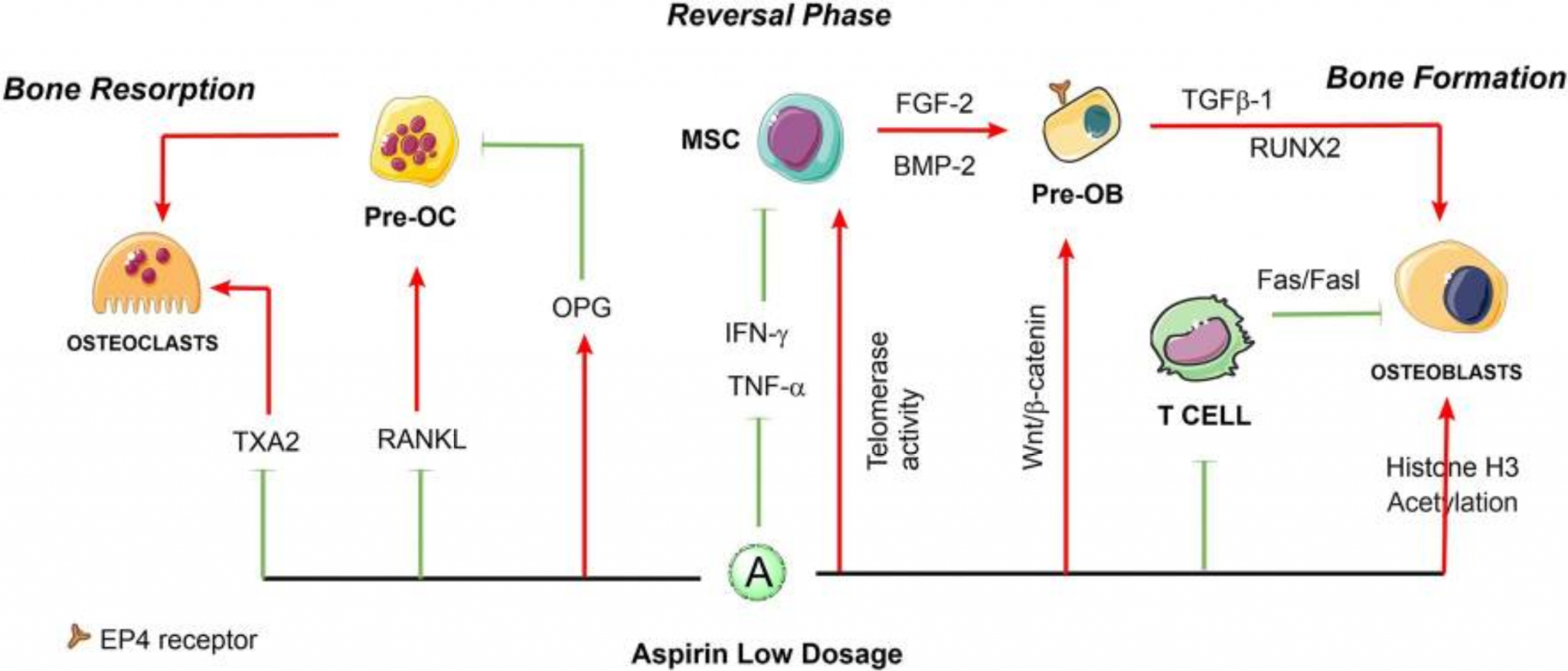
Low dose aspirin slightly upregulates telomerase activity in bone marrow mesenchymal stem cells (BMMSCs) and elongates their telomere lengths to avoid replicative senescence, improve stem cell functions, and has the capability to increase bone formation. 50μg/mL and 100μg/mL aspirin significantly increased transforming growth factor β-1 (TGF-β1) production of human BMMSCs, then induces migration of MSCs to the bone remodeling sites. Lower concentrations of aspirin (1μΜ and 10μM) promoted cell growth and increased ALP levels and RUNX2 expression, while higher concentrations (100μΜ and 1000μΜ) inhibited cell growth (P < 0.05), and lost their effect on ALP activity after 3 days, while even showing an inhibitory effect on the expression of RUNX2. Aspirin at a concentration of 100μM promoted cell mitosis from the S phase to the G2/M phase, and 1000μM arrested the cell cycle in the resting phase G0/G1 (P < 0.05). Parallel apoptosis/necrosis studies showed the percentage of cells in apoptosis decreased dramatically at any dose of aspirin. Mesenchymal stem cell-based intervention with low dose aspirin (< 100μg/mL) may benefit osteoporosis treatment by inhibition of osteoclast differentiation and activities. Aspirin at a dose of 50μg/mL can partially block the formation of osteoclasts induced by RANKL. The end result of these findings is that low dose aspirin promoted bone growth and high dose inhibits bone growth.
In the following schematic, low dose aspirin stimulates osteoblasts and inhibits osteoclasts. While there are not enough human clinical trials on low dose aspirin to establish low dose aspirin as a significant benefit for bone regeneration, there is no question from the pharmacology and animal studies that low does aspirin has positive effects on bone regeneration.
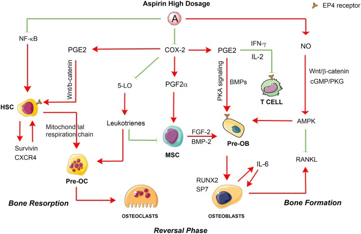
The red arrows indicate the promotion of cellular processes, and the green lines indicate inhibition of cellular processes.
The following schematic outlines the metabolic pathways altered by high dose aspirin showing an increase in bone resorption and a decrease in bone formation.
High Dose Aspirin (325mg) and Other NSAIDs
High dose aspirin and the other NSAIDs have similar metabolic pathways. While low dose aspirin (81mg) has an undeniable positive effect on bone formation and fracture healing, high dose aspirin and other NSAIDs have the opposite effect. In evaluating the pharmacology of high dose aspirin and other NSAIDs, there is a finding that the pharmacology of these medications indicates that they impart a negative effect on bone growth. In general, terms the overall effects of these medications inhibit osteoblasts and stimulate osteoclasts. However, there is considerable debate as to the clinical relevance of the negative pharmacology of these drugs when it come to fracture healing and bone regeneration.
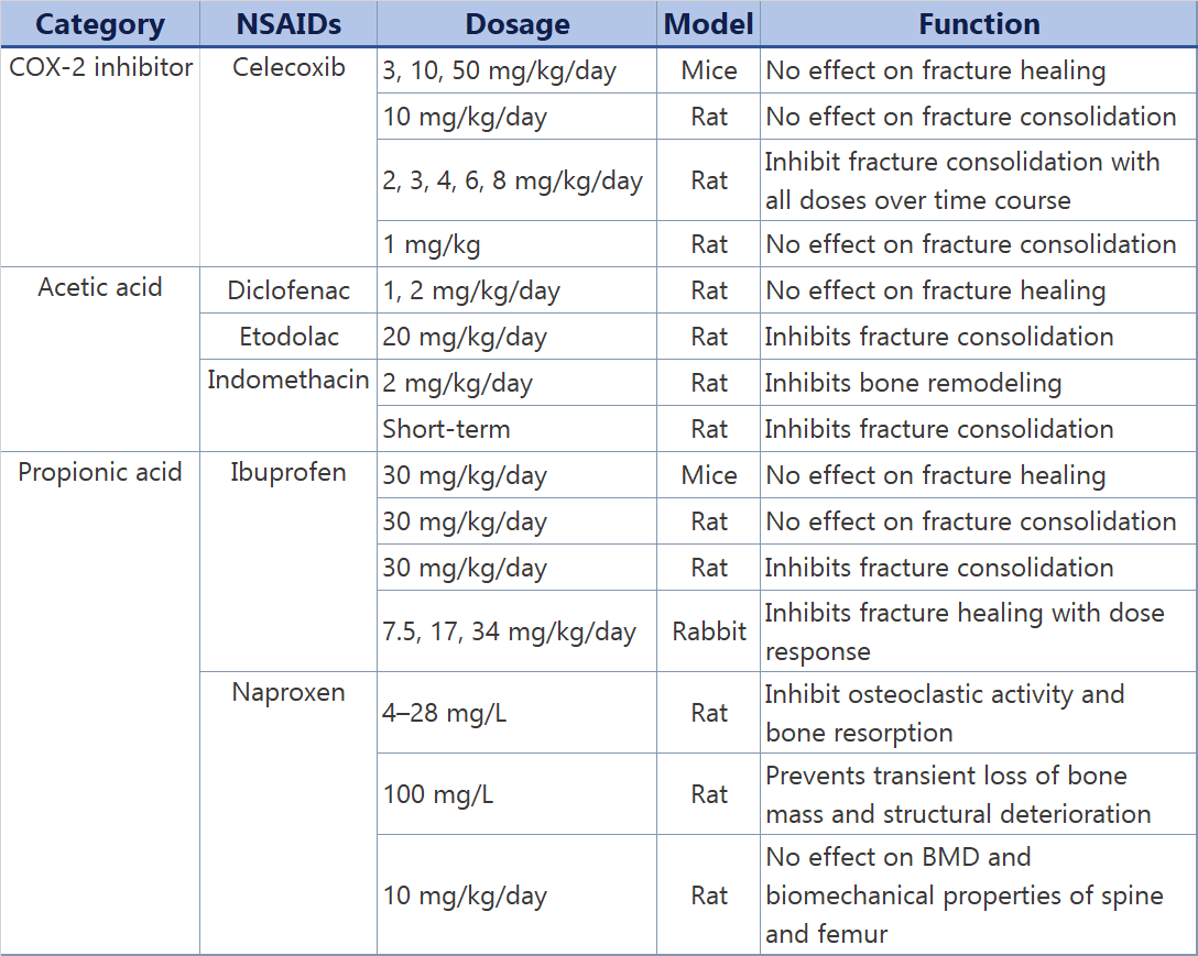
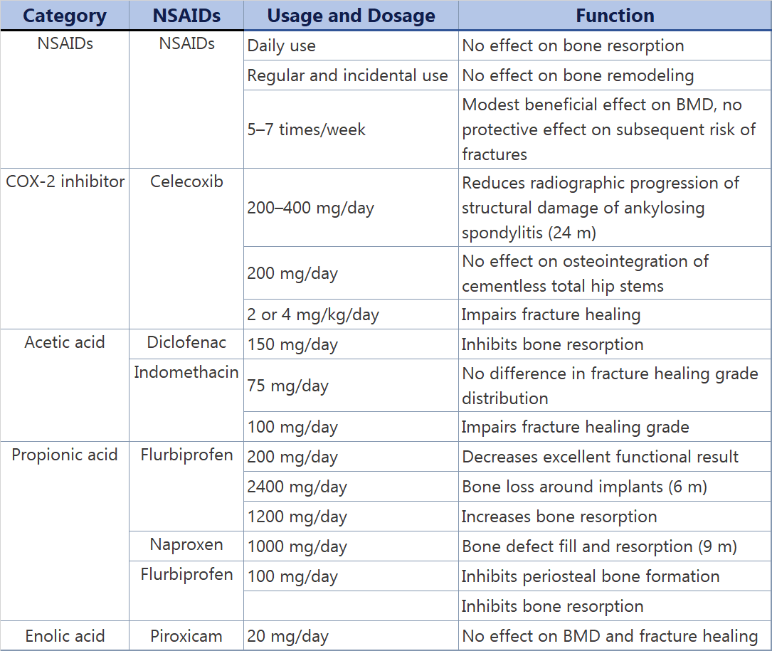
The two following charts summarize the animal and human clinical research on the effects of high dose aspirin and other NSAIDs used for pain control.
Table 1
Animal studies regarding the functions of NSAIDs in bone remodeling and fracture healing
To summarize these two tables, it can be seen that the studies found either no negative effect or an inhibitory effect on bone formation. But what about dental implants? One retrospective study evaluated long term implant failure in relation to NSAID consumption. One hundred and four patients with initially 468 implants had experienced 238 implant failures, of which 197 were due to failing osseointegration (42%). Sixty of the participants, initially with 273 implants, had used NSAIDs perioperatively and experienced 44% implant failures, versus 38% in the non-NSAID cohort. The NSAID cohort experienced 3.2 times more cases of radiographic bone loss greater than 30% of the vertical height of their remaining implants and 1.9 times more cases of cluster failures, defined as failure of 50% or more of the implant(s) placed. This retrospective study indicates that NSAID medications did result in a higher failure rate over time, but the nature of the study does not allow us to conclude that NSAIDs result in a higher implant failure rate.
Two studies done by the same group looked at bone to implant contact around titanium implants placed in rats. Click on each link to see the studies: