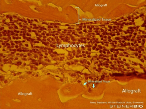
Normal bone formation:
- Vesicle budding: Osteoblast membranes bleb off 50–200 nm matrix vesicles (MVs) into newly laid osteoid near the cell surface.
- Ionic priming: MVs are enriched in alkaline phosphatase (TNAP) and PHOSPHO1, which raise local phosphate by hydrolyzing PPi and phosphoesters; annexins form Ca²⁺ channels; acidic phospholipids bind Ca²⁺.
- Nucleation inside the vesicle: First apatite nanocrystals appear within MVs.
- Vesicle rupture and spread: Growing crystals pierce the MV membrane, forming globular mineral nodules (“calcospherites”) that expand into the osteoid.
- Coalescence with the front: Nodules enlarge and eventually merge with each other and with the linear osteoid-front mineralization, after which lamellar maturation proceeds.
What you’d see:
- Light microscopy (H&E): scant or absent osteoid seams may hide it; when visible, look for patchy, globular basophilic foci within osteoid rather than a smooth linear front.
When it dominates:
- Very early bone formation / woven bone (fracture callus, early socket healing): Think of pericellular / matrix-vesicle–mediated mineralization as the kick start for normal mineralization. When initial normal bone formation begins, this method beings to mineralize the early collagen produced by the osteoblast, but soon extracellular ostoid-front takes over. Under normal conditions this process is not identifiable histologically.
Pericellular / matrix-vesicle–mediated mineralization does become an obvious, histologically identifiable entity in pathologic mineralization. When significant inflammation is present macrophages replace osteoblasts and are stimulated to become mineralizing cells that produce a histologically identifiable “rind” that covers inflammatory graft materials. This form of mineralization produces a mineral layer over cadaver graft particles. When the particles are covered by this rind, inflammation subsides in the surrounding tissue then the osteoblasts arrive to form dense sclerotic lamellar non-resorbable bone.
Example: The blue arrows are pointing at two cells producing pericellular / matrix-vesicle–mediated mineralization in the presence of inflammation. The process produces an acellular layer over the graft particle. In the following image this acellular layer is seen in the image as mineralized tissue. It is commonly called a rind.