BioDensification:

A Solution for Spinners, Immediate Implants, Sinus Grafts, and Soft Bone

It is not uncommon for an implant to lose bone at the apex within the sinus over time or for sinus augmentations to fail to regenerate sufficient bone to cover the apex of the implant. The following case is a method of regenerating the bone in the maxillary sinus for implant placement while at the same time covering the apex of a preexisting implant.

After the introduction of BioDensification for improving bone quality, a number of additional clinical uses have been developed, such as predictable implant integration for spinners, sinus augmentation without membrane exposure, and grafting immediate implants prior to implant placement.
BioDensificaiton for Increasing Bone Density and Vitality

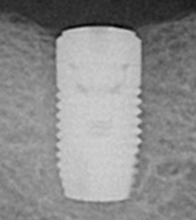
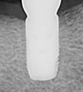
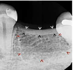
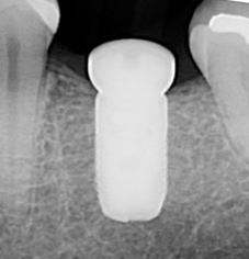
The pre-op image below shows poor mineralization under the crest. The osteotomy was prepared and filled with BioDensification prior to implant placement. Post-op radiograph is 3 months after grafting and implant placement. The crestal bone has remodeled and the implant is surrounded by dense normal trabecular bone.

Spinners

Spinners are a common problem in implant dentistry. The following pre-op radiograph shows an implant with no torque. The implant was retrieved, the socket filled with BioDensification, and the implant was replaced. The post-op radiograph is 3-months post implant placement and grafting shows excellent implant integration.

Immediate Implants

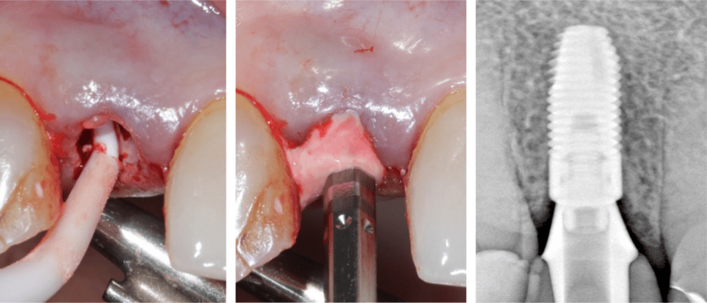
After extraction and osteotomy preparation, the socket is filled with BioDensification. Then, the implant is placed. BioDensifiaction is the only graft material proven to produce integration to the implant surface in the area of the graft. The result is a significant reduction in post op angular defects and improved long term success rates.

Trans-Osseous Sinus Augmentation

BioDensification’s ability to flow through bone allowed us to develop the most predictable and stress-free sinus augmentation procedure available today.

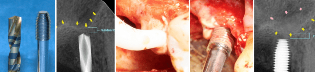
From the left, the Trans-Osseous Sinus Augmentation (TOSA) Kit consists of a specialized drill and driver. The osteotomy is prepared 1 to 2 mm short of the sinus as in the first radiograph. The next step is to fill the osteotomy with BioDensification as in the first clinical image. The second clinical image shows the driver placed in the osteotomy, where BioDensification is pressed into the bone. BioDensification flows through the bone lifting the sinus membrane off the floor of the sinus. If only a sinus bump is needed, the implant is then placed. If more lift is needed, additional graft material is used.

All SteinerBio bone grafts are fully resorbed, and produce normal healthy vital bone with proven long term success in grafted extraction sockets (3 years 100% integration 100% success). No other graft material matches this performance, making SteinerBio bone grafts the only bone grafts cleared by the FDA for implant placement.

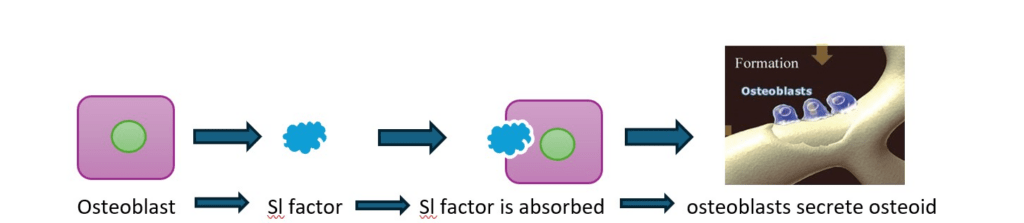
To understand why, here is a simplified schematic of the biological activity unique to SteinerBio bone grafts:

Osteogenic stimulation occurs as osteoblasts secret osteoid, the first layer of bone formation. During this process, the stimulated osteoblasts migrate, increasing bone density, integrating to the implant surface as your patient produces their own bone that is stronger and healthier.

MEMBER:

American Society for Bone and Mineral Research (ASBMR)

Tissue Engineering and Regenerative Medicine International Society (TERMIS)

American Academy of Implant Dentistry (AAID)