
Here’s a compact, end-to-end view of how bone grafts are resorbed—who the cells are, how they form, and how they actually digest the material. I’ll separate cell biology from chemistry at the interface and call out what differs across graft types (βTCPvs HA/xenograft/allograft).
Q. Who does the resorbing?
A. Osteoclast lineage (specialized bone-resorbing cells)
- Origin: circulating monocytes (hematopoietic).
- Commitment & fusion:
- M-CSF (macrophage colony-stimulating factor) → survival/proliferation of precursors.
- RANKL (from osteoblasts, stromal cells, activated T cells) + RANK on precursors → master transcription factor NFATc1 → osteoclast program.
- Cell–cell fusion via DC-STAMP/OC-STAMP, with co-receptors (Dap12, c-Fos), yields multinucleated osteoclasts.
- Attachment: αvβ3 integrin on the osteoclast binds the osteoclast to the Beta TCP granule creating a tight sealing zone (actin ring).
Q. How the resorption apparatus digest a particle?
A. Osteoclast “acid + enzyme” system (on a sealed lacuna)
- Polarization & sealing
- Actin ring → sealing zone; inside it forms the ruffled border (secretory membrane).
- Acidification (demineralization step)
- Carbonic anhydrase II makes H⁺ from CO₂ + H₂O.
- V-ATPase pumps H⁺ into the lacuna to pH ~4.3–4.7.
- ClC-7 (chloride channel) balances charge.
- Result: apatite dissolves → Ca²⁺ and PO₄³⁻ go into solution.
- Transcytosis / cleanup
- Solubilized ions and peptides are endocytosed at ruffled border and released at the basolateral membrane; some calcium/phosphate can be reused locally.
Caveat: Not all beta TCP is the same. The performance of a beta TCP is directly related to its structure, hardness, and purity. The image below is an XRD of our OssseoConduct βTCP graft material.
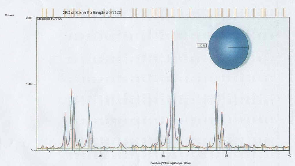
This XRD shows OsseoConduct is a 100% pure beta TCP with a perfect crystalline structure and no contaminants.
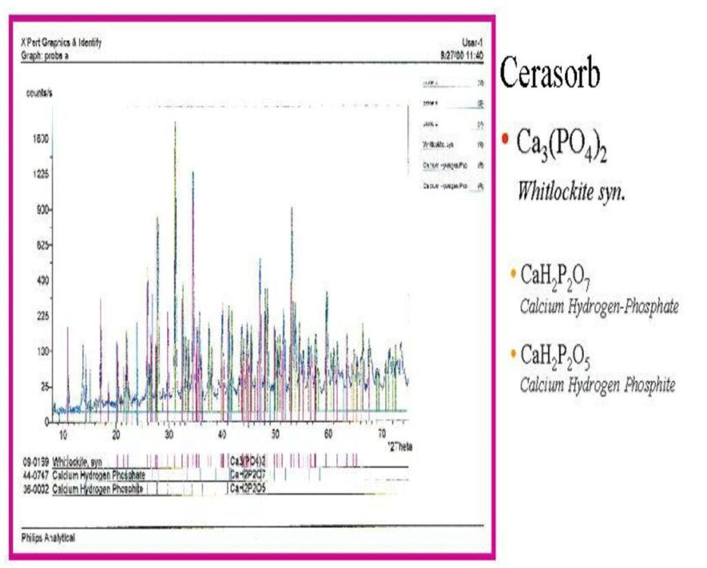
Pyrophosphate (PPi) is a potent, natural inhibitor of mineralization. If PPi-bearing salts contaminate a βTCP/whitlockite graft, they can measurably depress new-bone formation until they’re hydrolyzed to orthophosphate.
What PPi does in bone formation:
- Blocks crystal growth: PPi (P–O–P bond) adsorbs to nascent apatite surfaces and prevents further Ca–P deposition. Osteoblasts normally counter this by alkaline phosphatase (ALP), which cleaves PPi → 2 Pi, removing the brake.
- Ties up free Ca²⁺: PPi complexes Ca²⁺, lowering supersaturation for apatite.
- Local acidity (for acidic salts): Acid calcium pyrophosphate (e.g., CaH₂P₂O₇) can transiently acidify microenvironments as it dissolves, further disfavoring apatite precipitation.
Practical impact on a bone graft:
- Slower osteoid mineralization → delayed radiopacity.
- Lower early bone volume fraction at 2–8 weeks vs. a clean β-TCP/HA control.
- More fibrous tissue persistence in spaces that would otherwise mineralize.
- Pro-inflammatory risk if crystals shed: poorly controlled CPP phases can release calcium pyrophosphate crystals (biologically similar to those in CPPD/pseudogout) that may provoke macrophage/neutrophil responses, usually mild in dental defects but still undesirable.
- Remodeling lag, because resorption–reprecipitation cycles depend on available orthophosphate.
Which contaminants matter here:
- CaH₂P₂O₇ (acid calcium pyrophosphate): most concerning; more soluble → more PPi in the wound early on.
- Ca₂P₂O₇ (neutral calcium pyrophosphate): less soluble; still inhibitory where particles contact osteoid.
- Any mixture showing the PPi “signature” will act similarly until ALP (and time) convert PPi to Pi.
