Pre-operative Antibiotics for Extractions

Two studies published in the Journal of Bone and Mineral Research found that tooth associated infections such as periodontitis and endodontic pathology affect the ability of the socket to heal after extraction.

With the understanding that infection associated with a tooth compromises the ability of the socket to heal, SteinerBio sought to find a way to improve socket healing after extractions when significant infection is present. Our thought was to heal the bone before we removed the tooth. The method employed was to prescribe Amoxicillin 500 mg TID 3 days prior to tooth extraction followed by 7 days post-extraction. It is also our experience that when a maxillary posterior tooth is severely infected, this commonly involved the sinus resulting in even more compromised healing of these extraction sockets. In cases of significant infection of maxillary posterior teeth, the antibiotic protocol was Augmentin 500 mg 3 days prior to extraction and 7 days post extraction.
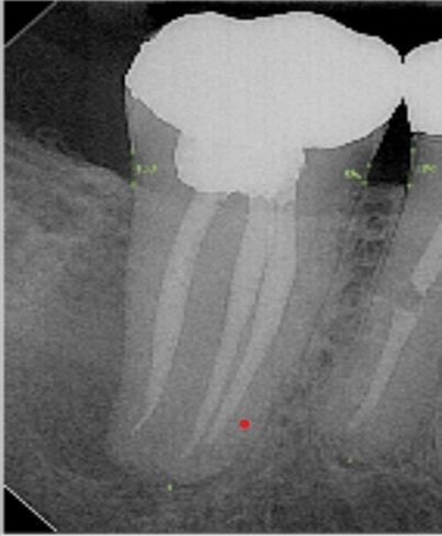
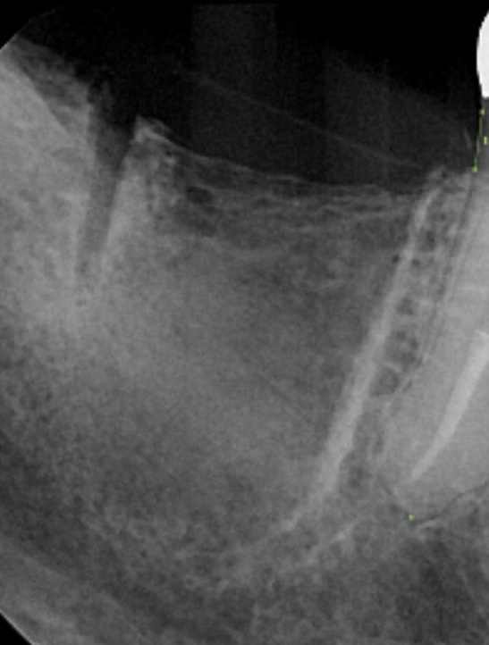
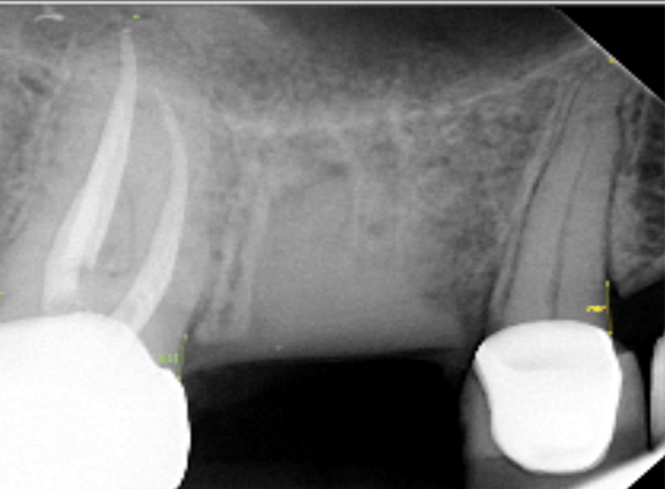
Pre-op with significant bone loss

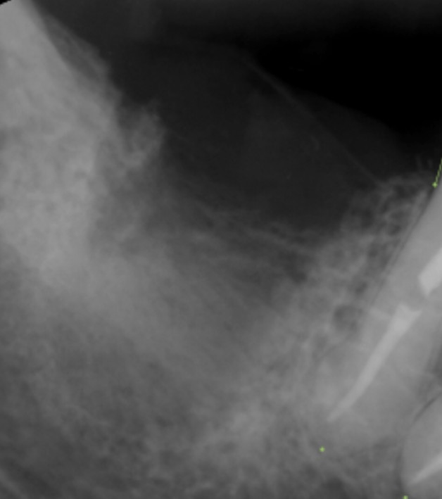
Day of extraction and grafting with Socket Graft Injectable. Covered with a d-PTFE membrane.

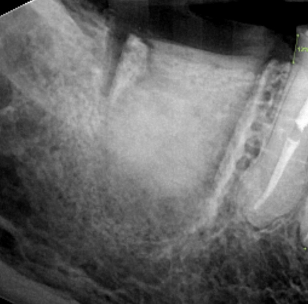
Two-week post-op. At this time period mineralization is beginning in the apex of the extraction socket. In the coronal portion of the socket the graft material remains but it is much more radioleucent than when the graft was placed two weeks prior. When the graft material is placed extracellular fluid washes out the soluble portion of the graft material that makes the remaining graft material porous allowing for migration of regenerate cells into the graft material. In this image you are looking at residual graft material in the coronal portion of the extraction socket.

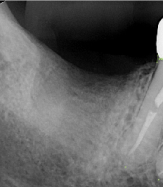
One-month post-op. The membrane is removed. Mineralization has displaced the majority of the graft material which is no longer visible in the radiograph. Mineralization is visible in 80% of the socket. Mineralization is intense at this time frame with the highest number of osteoblasts actively forming bone.

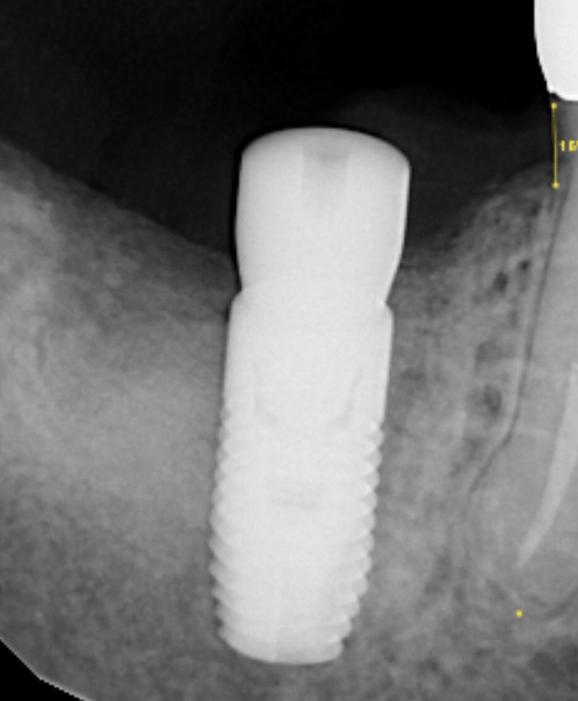
3-month post-op for implant placement. Mineralization in the body of the socket has now equaled the mineral content of the surrounding bone. The crest is well mineralized.

At 3-months post extraction and graft, the implant is placed in healthy vital bone. No graft material is remaining at this time frame.

The purpose for using this antibiotic protocol was to improve the regenerative process in the presence of significant infection. The results have been very positive, producing excellent rapid bone regeneration and ridge maintenance.

However, another significant benefit was noted during the extraction appointment. Along with reducing the inflammatory response to the infection in the bone, the granulation tissue was significantly reduce making tissue removal much easier and a significant reduction in bleeding as is noted in the following images.

In this image, there is significant interproximal bone loss. In addition, there is infection involving the sinus. This infection will compromise all of the surrounding bone with the addition of inflammatory tissue in the sinus.

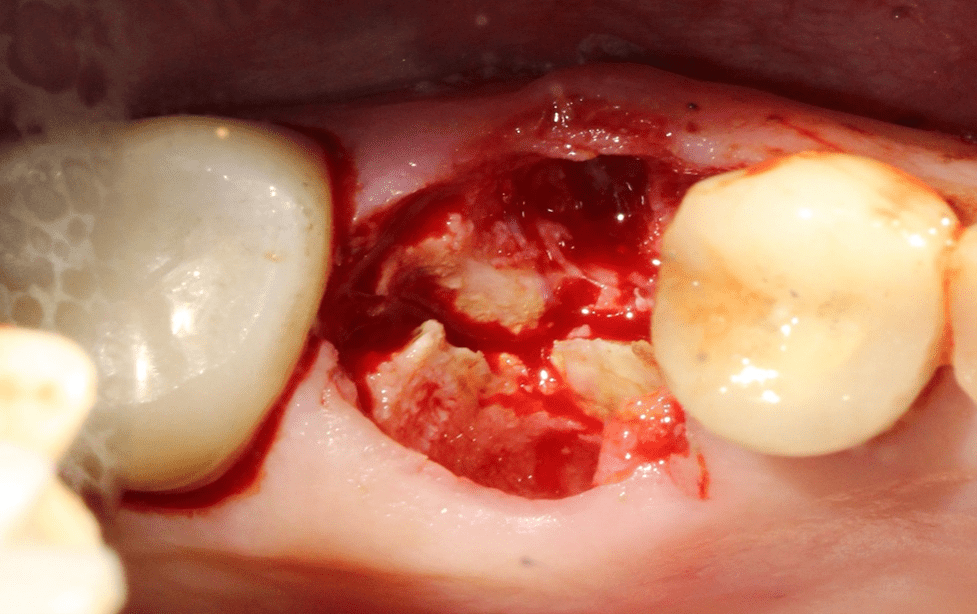
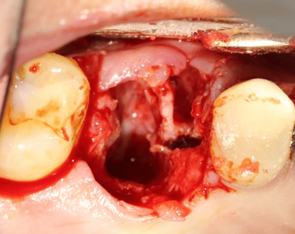
Normally, a clinician would expect significant granulation tissue and intense bleeding from the extraction socket. However, rather than encountering granulation tissue with significant bleeding, the tissue in the socket was more like fibrous tissue than hemorrhagic granulation tissue. The fibrous tissue is not firmly attached to the bone making removal of the fibrous tissue from the socket easy with virtually no bleeding.

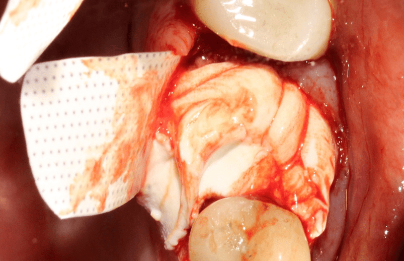
The membrane is prepared before grafting by creating 6mm pockets buccally and lingually. Socket Graft Injectable is injected into the socket and covered with a membrane. Socket Graft Injectable does not require packing. Inject and cover.

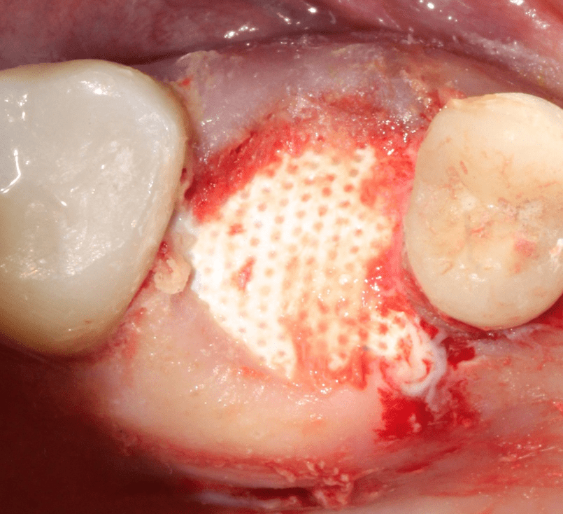
After tucking the membrane, Oral Bond is applied onto the membrane creating a hermetic seal. No sutures are required.

Day of surgery with graft in place

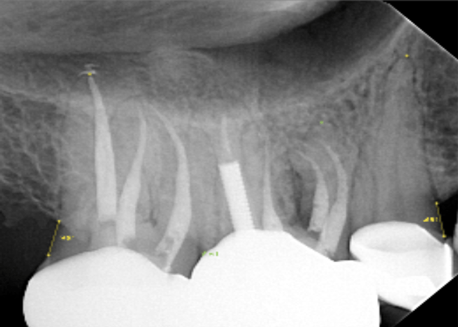
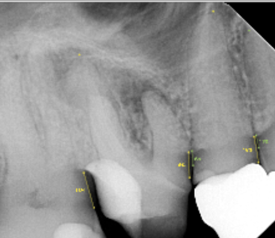
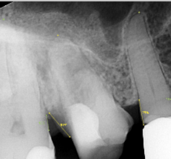
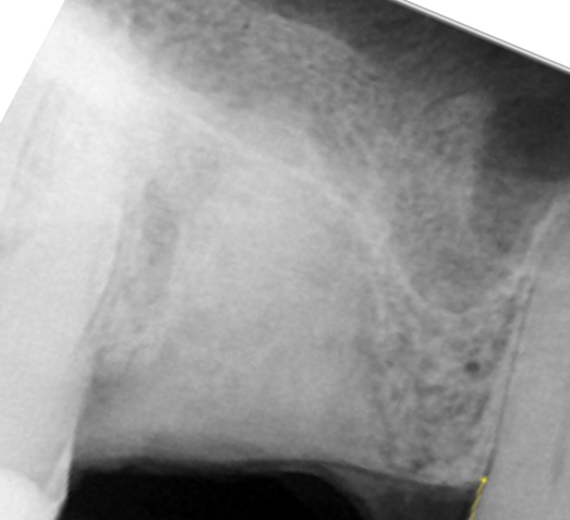
This is another case. In this radiograph, significant bone loss is noted with significant sinus involvement 5 months prior to the extraction.

The patient was prescribed Augmentin 500 mg 3 days prior to this radiograph taken on the day of the extraction.

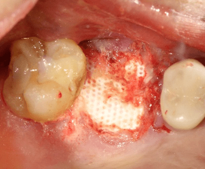
After 3 days of Augmentin, the socket presented with no granulation tissue. The socket was filled with fibrous tissue that was easily removed. Nothing was done to the socket except to curette out the fibrous tissue. Minimal bleeding was noted. As with the previous case, we injected Socket Graft Injectable and covered with a d-PTFE membrane and Oral Bond. No packing and no sutures.

Day of extraction and grafting with Socket Graft Injectable.

Extraction socket is covered with d-PTFE membrane sealed with Oral Bond only and no sutures.

This suture-less membrane technique allows the tissues to heal freely without constraining and collapsing the gingiva in addition the patients don’t have to deal with sutures.

In cases where there is significant infection from either periodontal disease or endodontic disease, 3 days of antibiotics prior to the extraction appears to significantly improve bone regeneration and ridge maintenance. In addition to improving bone regeneration, removal of residual tissue in the socket is made easier as the tissue is not firmly bound to the bone as is found with normal granulation tissue. After removal of the fibrous tissue, very little bleeding is experienced facilitating graft placement and retention. In these cases all 4 bony walls were present. When the socket walls are damaged we advise using Socket Graft Plus for regeneration of the socket walls and sutures to stabilize the graft material. At SteinerBio, pre-op extraction antibiotics for infected sockets is now routine protocol for successful bone regeneration.

MEMBER:

American Society for Bone and Mineral Research (ASBMR)

Tissue Engineering and Regenerative Medicine International Society (TERMIS)

American Academy of Implant Dentistry (AAID)