Learning the Inverted Periosteal Graft
This patient is a healthy middle aged black male. The treatment was the inverted periosteal graft using Immediate Graft mixed with OsseoConduct βTCP granules as graft material.
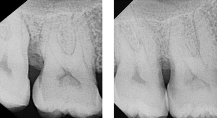
Left: Pre op radiograph with 8mm probing
Right: 6 week post op with 3mm probing
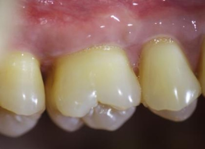
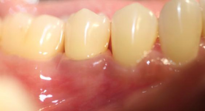
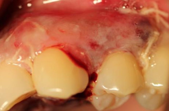
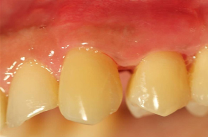
6-week post op photograph buccal
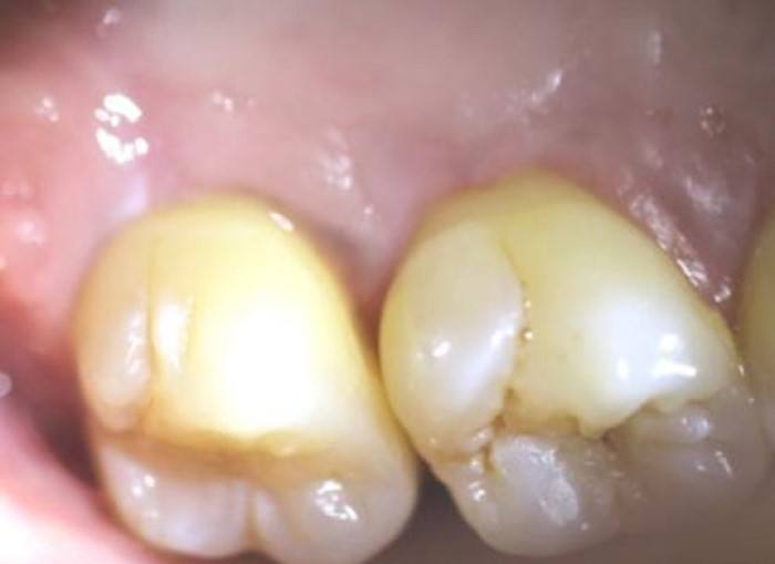
6-week post op palatal
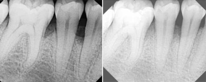
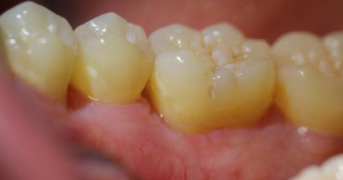
6-week post op photograph lower right buccal
Post op photograph, LR lingual
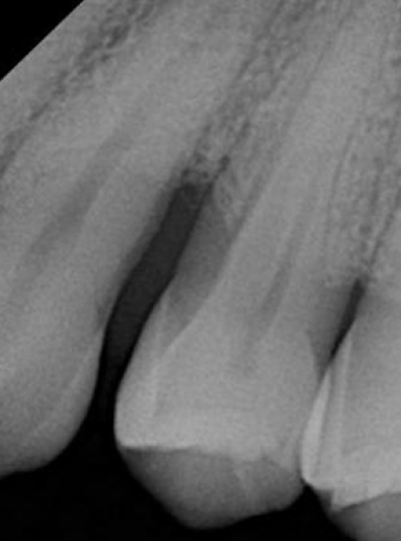
Pre op radiograph with probing mesial #12, 8mm
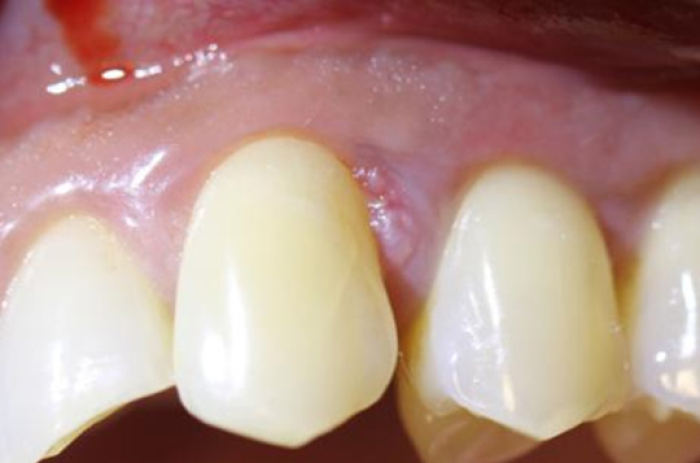
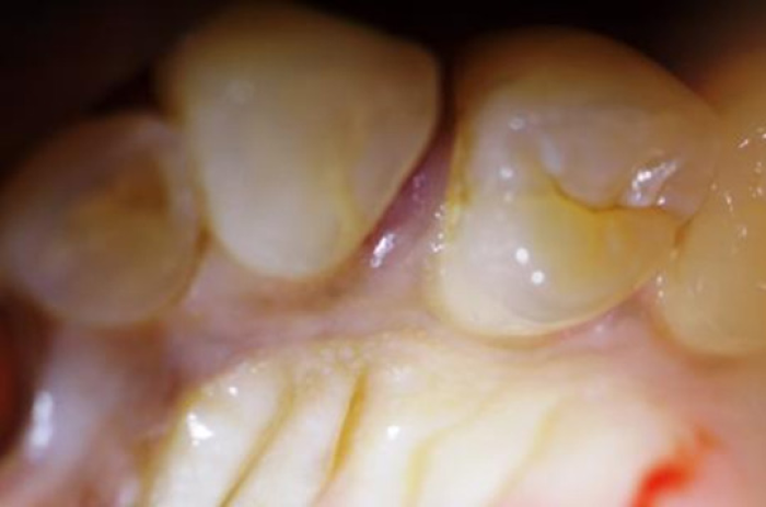
Pre op photograph palatal
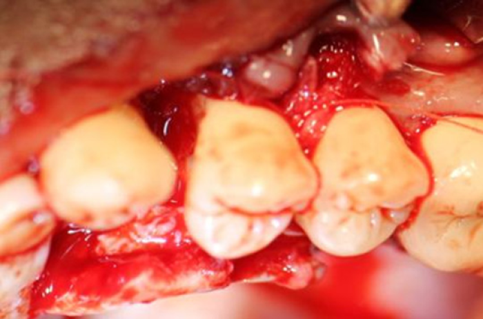
Vertical releasing incisions are made one tooth mesial and distal to the area to be regenerated. Sulcular incisions are used with no scalloping.
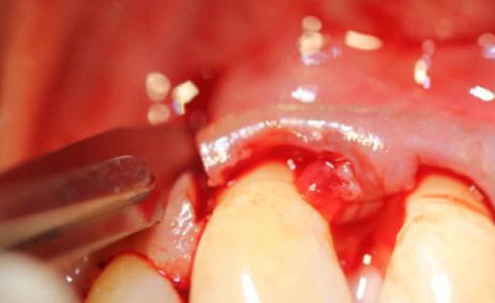
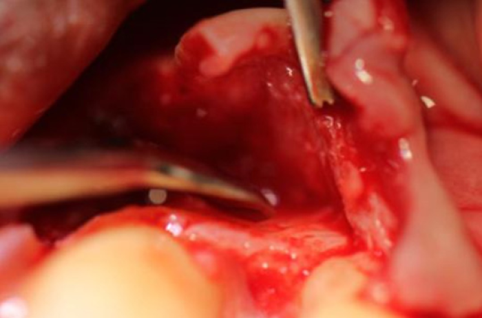
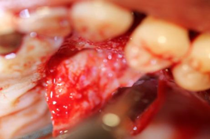
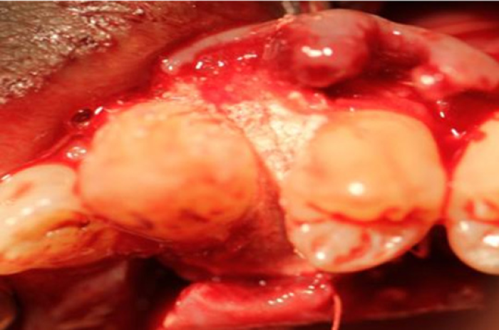
The gingiva is reflected to the mucogingival junction
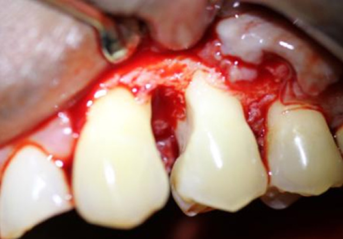
The roots are planed and conditioned with PrefGel
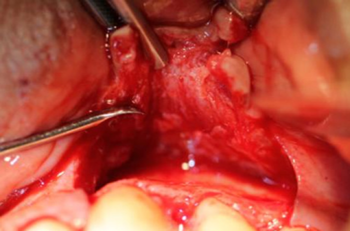
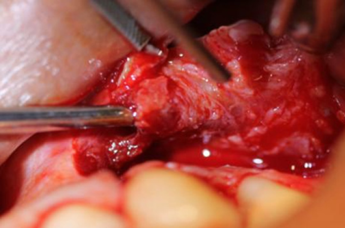
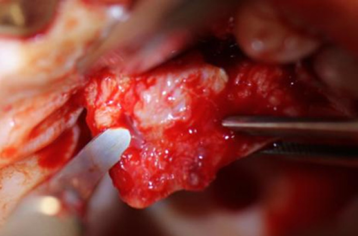
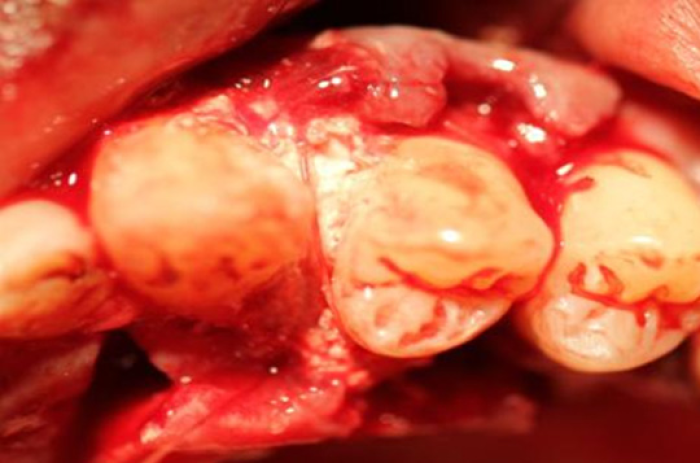
The periosteum is dissected from the alveolus cleanly with a sharp spoon.
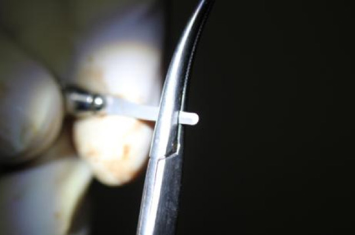
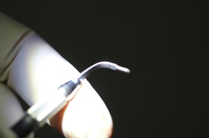
A small angled spoon is used to locate the edge of the periosteum.
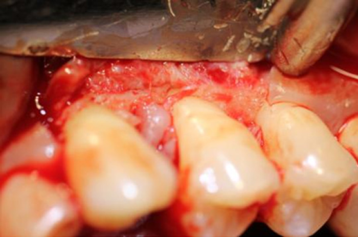
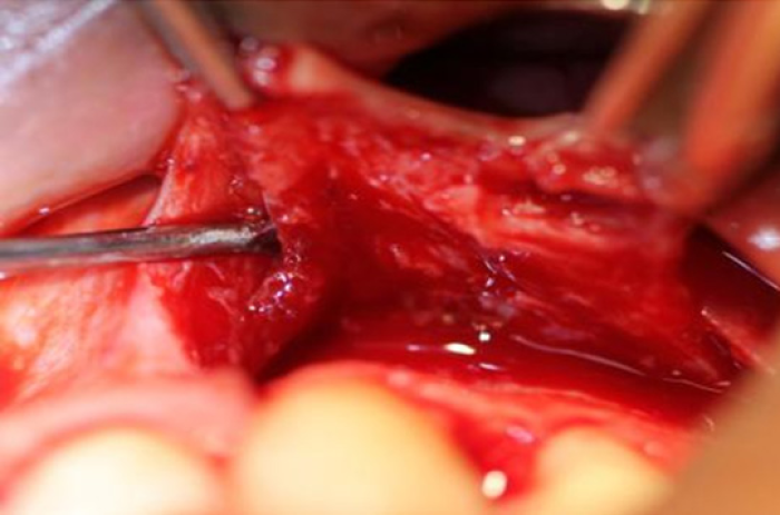
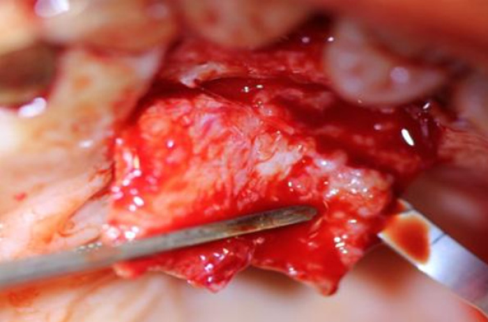
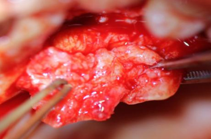
The small spoon is inserted under the periosteum. The periosteum is dissected off the buccal flap from the mucogingival junction to the base of the flap along the full length of the flap.
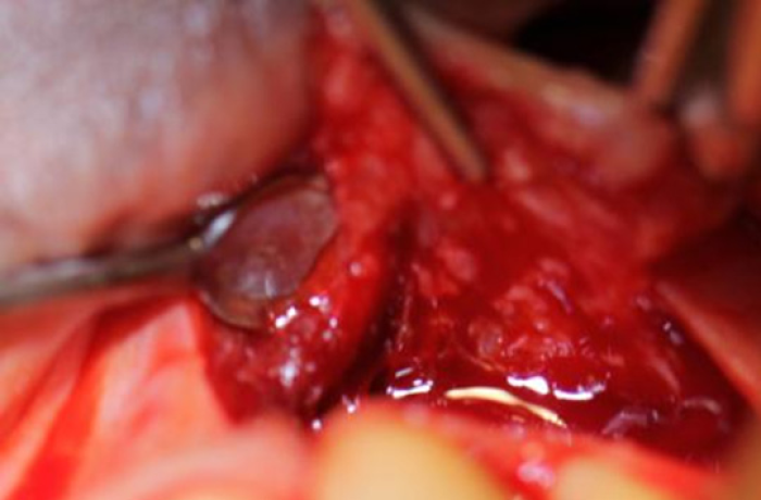
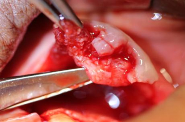
After the dissection with the small spoon, a large spoon is used to complete the dissection.
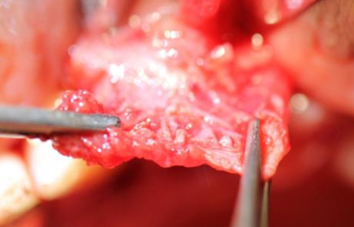
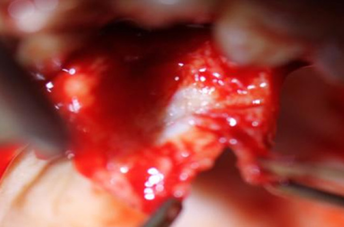
The large spoon is completes the dissection throughout the full length of the flap.
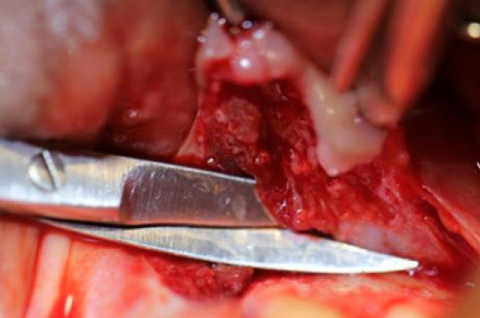
La Grange scissors are used to cut the periosteum at the base of the flap.
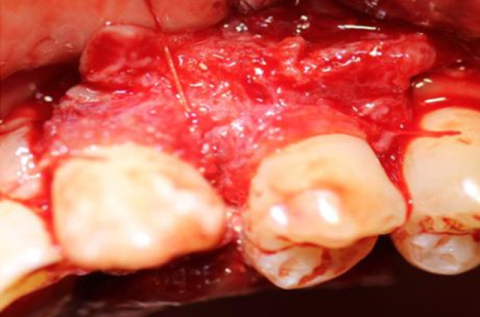
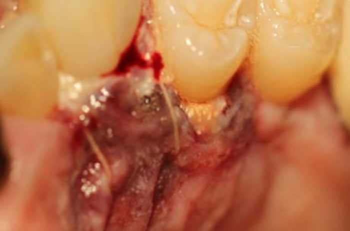
The suture is tied drawing the periosteum completely over the graft, resulting in the buccal and lingual periosteum to connect interproximally. Note that in this case the periosteum was sutured both mesial and distal before closing over the graft. When the periosteum is closed, the flaps are sutured.
The inverted periosteal graft places regenerative cells over the area to be regenerated. In addition, the periosteum is an ideal barrier to unwanted cells. The attached gingiva and the periosteum will not tolerate contact with each other and therefore the periosteum is an ideal biological barrier. In the posterior, the papilla will not lay over the periosteum. In the anterior, the papilla will lay over the periosteum.
A minimum of 6 weeks is required before the tissues can reorganize and the periodontal ligament can be probed. Over a few months, the tissue will fully reorganize into normal anatomy. The radiographic appearance of the bone will continue to increase in radiodensity over the following months and a periodontal ligament will appear radiographically.
This surgery is very technique sensitive. The methods and materials have been developed over a 10-year period and any alteration in technique or materials will likely lead to failure of this surgery. It is advised that the surgeon follow instructions precisely until experience is gained.