Clinical Outcomes of Graft Selection on Final Bone Quality

The purpose of this paper is to give you an understanding of bone formation at a tissue level. We will be looking at cells and the structures that those cells are creating so you can see in your mind what you are creating in your patients. Just as importantly, you will be able to direct the process of bone formation that you want for your patient by the materials you use.

While this paper focuses on the tissue level of bone regeneration, we will also present sections that are labeled DEEP DIVE. The deep dive buttons are links to what is going on physiologically inside the cell and discuss the cellular mechanism responsible for the tissue you are seeing in the histology. We suggest that you read the paper and get an understanding of the tissues you are creating and only if you are interested you can go back to DEEP DIVE for the mechanisms of osteogenesis.

First, it’s important to understand some terms for clarity of the topic. We will discuss the scientific terms that are used to describe how bone forms in your patients and then we will look at the histology that describes each of these processes. Let’s get started:

Terms

  • Osteogenesis: the entire bone‐forming program from stem/progenitor stimulation and commitment → osteoblast differentiation → osteoid deposition → mineralization → lifelong remodeling/repair.
  • Intramembranous osteogenesis: osteoblasts lay down osteoid and then mineralize it; referring to the type of bone formation in the jaws, skull, and clavicle.
  • Endochondral osteogenesis: cartilage is formed and then replaced by bone. This refers to bone formation in the rest of the skeleton, and we will not discuss this form of osteogenesis in this paper.
  • De Novo intragraft osteogenesis: where bony islands start inside a graft matrix. This type of osteogenesis occurs when bone producing cells crawl into a matrix and begin to randomly form bone in the matrix without being on a surface.

Terms that describe where the bone is being produced:

  • Distance osteogenesis: Bone grows from the surrounding bone and fills the bony defect.
  • Contact osteogenesis: where bone starts on a material surface; This is the type of osteogenesis when bone first forms on an implant surface.

Just to confuse you more, we also have different types of mineralization. Mineralization is the phase of osteogenesis that ossifies and hardens the bone tissue.

  • Mineralization (biomineralization): deposition of any inorganic mineral in tissue. It can be physiologic (bone, dentin, enamel) or pathologic (atherosclerosis, the mineralization of damaged tissue via trauma or the production of sclerotic bone around inflammatory grafts). Mineralization is always produced by cells. There are two types of mineralization: osteoid-front mineralization and pericellular / matrix-vesicle–mediated mineralization.
    • Osteoid-front mineralization: the advancing interface where osteoblasts produce unmineralized osteoid that becomes mineralized bone. Think of a moving boundary right under a row of active osteoblasts.
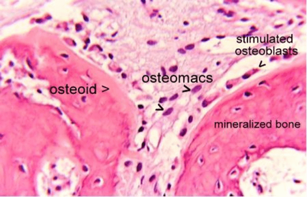
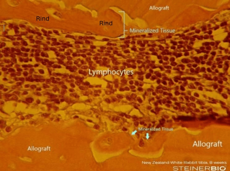
Example of osteoid-front mineralization:

What you see histologically:

  • Osteoblast rim: plump, cuboidal cells lining the surface
  • Osteoid seam: pale eosinophilic band just beneath the osteoblasts
  • Mineralization front: a thin basophilic line where osteoid transitions to mineralized bone

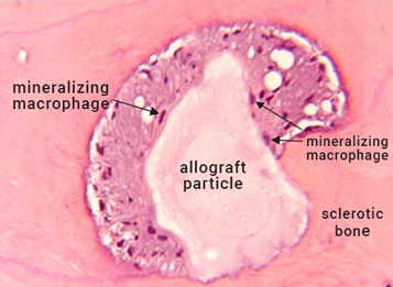
Pericellular / matrix-vesicle–mediated mineralization: is the nodular (calcospheritic) start of mineral deposition that originates in extracellular vesicles budded from mineral forming cells, mainly osteoblasts (also chondrocytes in cartilage, odontoblasts in dentin and inflammation induced mineralizing macrophages).

  • Physiologic osteoid: Pericellular matrix-vesicle -mediated nucleation is classically from osteoblast-derived vesicles (rich in TNAP, PHOSPHO1, annexins), seeding calcospherites that coalesce into a mineralization front. This is a kick-start to osteoid-front mineralization.
  • Inflamed fields: When there’s significant inflammation, matrix-vesicle -like particles that nucleate apatite are often myeloid-derived, and “mineralizing macrophages” are a common source. Macrophages (and foam cells) can express ALP, release annexin-positive extracellular vesicles, and drive dystrophic/ectopic calcification.
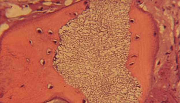
    • Example: the image below shows Pericellular / matrix-vesicle–mediated mineralization in an inflamed field.
  • Osteoconduction: is the process by which a graft acts as a scaffold that lets the patient’s own bone-forming cells grow onto it. Osteoconduction occurs via distance osteogenesis as bone grows in from the surrounding tissue. The graft material provides a biocompatible scaffold for cellular and vascular ingrowth. Distance osteogenesis always starts from preexisting bone and grows outward and when it meets an osteoconductive scaffold bone grows on the surface of the scaffold and integrates into the graft material.

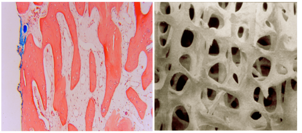
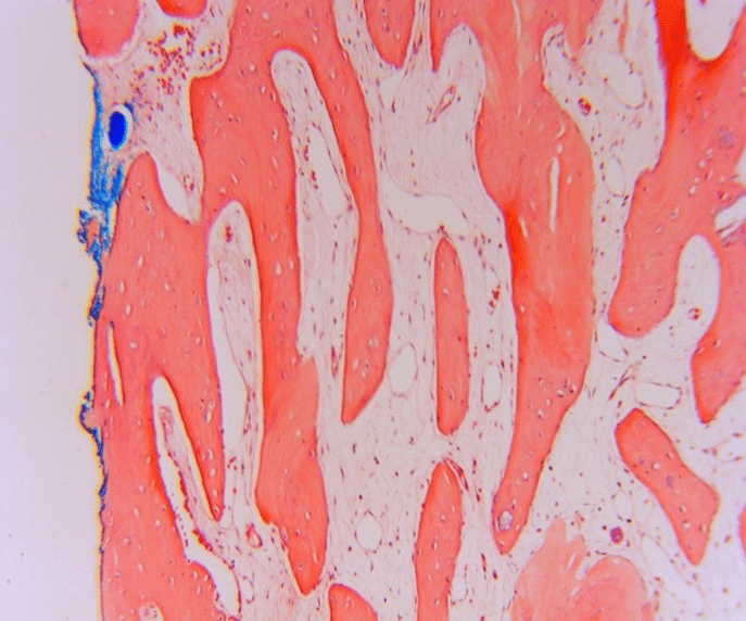
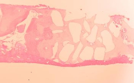
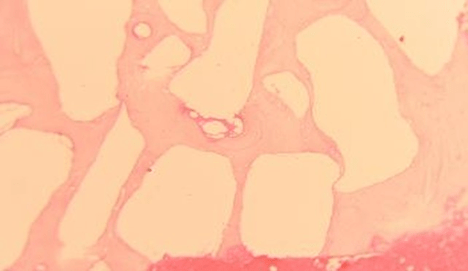
Before we begin, take a look at the two images above. These images are ideal trabecular bone that you want to put your implant in for a lifetime of function.

Natural Socket Healing

Distance Intramembranous osteogenesis with osteoid front mineralization:

This is a healing socket without grafting. The image shows bone growing in from the sides of the socket wall toward the center of the socket.

Type of osteogenesis:

  • Intramembranous Distance Osteogenesis
    • New bone is originating on the host socket walls and growing toward the center of the socket.

Form of mineralization:

  • Osteoid-front mineralization
    • Osteoblasts lay down osteoid on advancing fronts; mineral forms on the osteoid by heterogeneous nucleation and calcospheritic deposition within the osteoid, creating woven bone.

In short: a 5-week socket shows distance intramembranous osteogenesis with osteoid-front mineralization producing immature woven bone that is bridging inward from the native bony walls. A key feature is bone growing from the preexisting bone to fill the void.

.

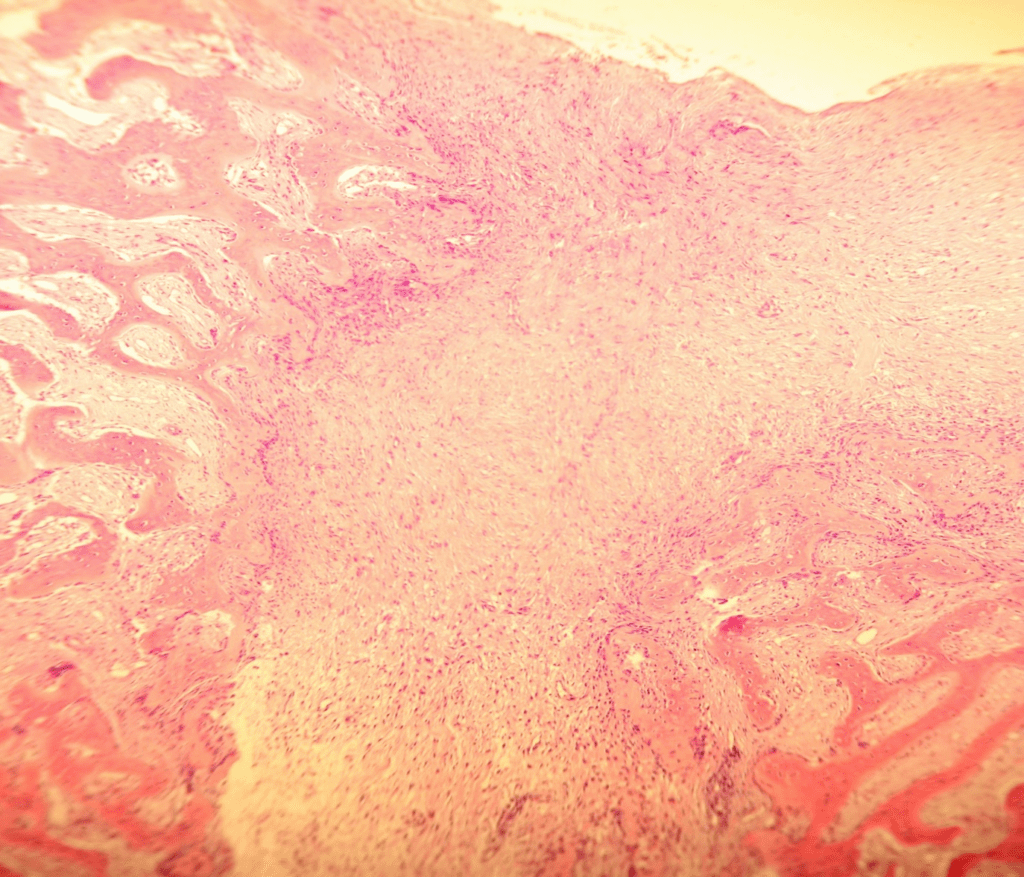
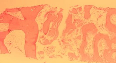
Here is a common result of socket healing with no graft. The trabeculae are weak and collapse when biopsied. This is the type of bone you experience when very little resistance to drilling is felt.

Clinical Significance: This process is bone repair that often produces good bone for implant integration. However, when bone heals unassisted it is never equal to normal healthy vital bone. The vitality of the repaired bony defect can be anywhere from good bone to very poor quality bone which will determine if your implant integrates and functions long term. In addition, the volume of unassisted repaired bone is always inferior due to resorption. 50% of extraction sites that heal in this manner require ridge augmentation for implant placement.

Resorbable Bone Graft Granules – Beta Tricalcium Phosphate (βTCP)

Distance Intramembranous osteogenesis with osteoconductive osteoid-front mineralization.

This image is a OsseoConduct βTCP granule 4 months after ridge augmentation. The bone has grown in from the surrounding bone (distance) and has grown over the graft particle (osteoconduction). This is distance osteogenesis from the surrounding bone with the biologic process of intramembranous osteogenesis producing osteoid-front mineralization.

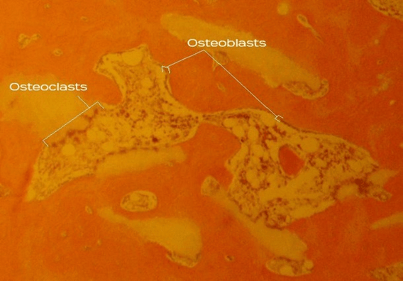
A lower power showing multiple OsseoConduct βTCP granules surrounded by both osteoclast resorption of the granules and osteoblasts that are involved in intramembranous osteogenesis and osteoid-front mineralization. This is bone remodeling from immature woven bone encasing OsseoConduct βTCP granules. As the granules are resorbed the woven bone is converted into ideal lamellar bone.

  • Osteoclasts (multinucleated, foamy cytoplasm) are sitting in Howship’s lacunae along the beta TCP surface, showing clear evidence of cell-mediated resorption of the granule.
  • Osteoblasts form a continuous lining on adjacent trabecular surfaces, laying down an eosinophilic osteoid seam that is mineralizing.
  • The trabeculae have the irregular texture of woven bone, with early osteocyte lacunae; you can also make out reversal/cement lines at some borders.

The end result of the normal remodeling shown with this material is the production of normal healthy vital bone as seen in the above image.

Clinical Significance: All of the processes shown in this form of bone creation are normal processes. The normal process of bone formation is modified such that that granules is left in place long enough to maintain volume until bone can fill the space. Once the area is filled with bone the OsseoConduct βTCP particles are resorbed leaving nothing but normal healthy vital bone. The end result is maintenance of the jaw volume due to osteoconduction of the graft material. The FDA has very stringent requirements for clearing a bone graft for implant placement. OsseoConduct βTCP granules is the only granular graft material cleared by the FDA for implant placement due to its ability to produce normal vital bone.

Distance intramembranous osteoid-front mineralization without osteoconduction.

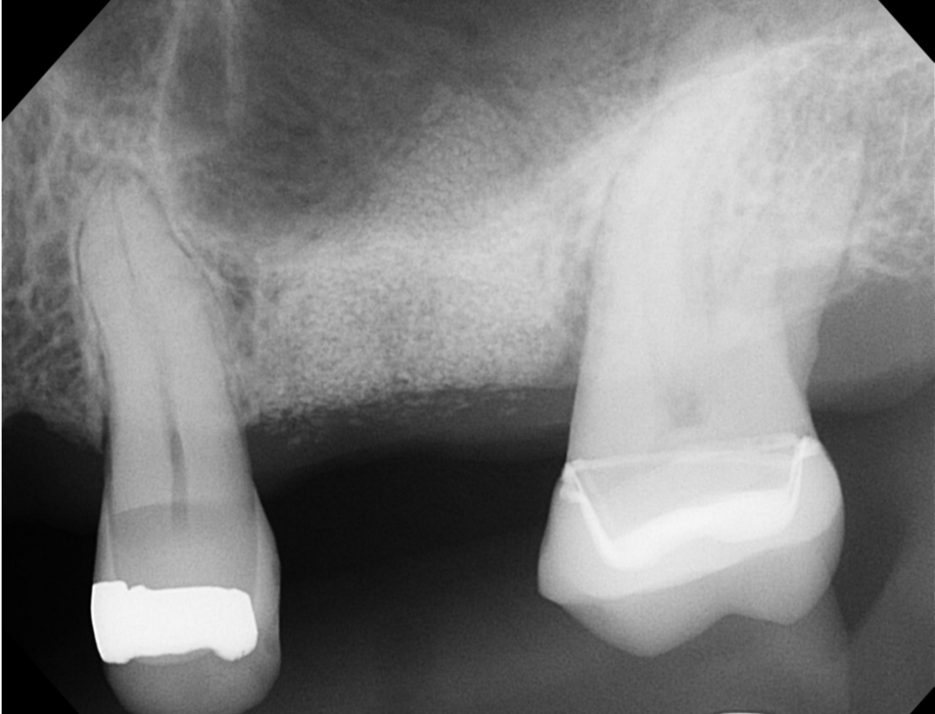
X-ray shows 6 months after grafting with Symbios by Dentsply

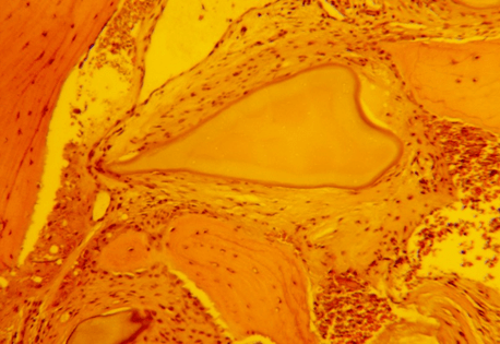
6-months post-op histological slide showing Symbios. The material is 80% beta TCP and 20% hydroxyapatite.

  • Central graft particle: pale, acellular body with smooth contours; still a non-vital residual granule.
  • Capsule: immediately surrounding it is fibrovascular connective tissue, not lamellar bone; wavy collagen bundles with spindle-shaped fibroblasts and small vessels; no osteocyte lacunae contiguous with the particle.
  • No bone contact: No osteoid seams, lining osteoblasts, or Howship’s lacunae at the interface; the border is relatively featureless; typical of fibrous encapsulation rather than replacement or active remodeling.
  • Adjacent bone (where present): separated from the particle by a fibrous band; any lamellar organization is outside that band, not touching the particle.
  • Inflammation: minimal; no conspicuous giant-cell reaction, so this looks like a quiescent fibrous capsule rather than an active foreign-body response.

Implication: This is a persistent graft particle isolated by a fibrous capsule; a pattern frequently reported with certain allograft/xenograft particles. Functionally, it means the particle isn’t integrated into load-bearing bone and contributes little to remodeling beyond space maintenance.

This is an example of distance osteogenesis without osteoconduction. The graft material is just being pushed aside as bone grows into the socket. The bone graft is taking up space and providing no support which will compromise the ability of the site to support integration and implant function. This material does not qualify to be considered a bone graft and should not be used in areas that are intended for implant placement.

Non-Resorbable Bone Graft Granules – Hydroxyapatite (HA)

Distance Intramembranous Osteogenesis with osteoid–front mineralization with osteoconduction without remodeling:

Low-power histology of a grafted extraction socket

  • Numerous pale, smooth-edged, polygonal/ovoid islands are present; these are cross-sections of non-resorbable hydroxyapatite (HA) granules
  • Around and between the granules is a dense, uniformly eosinophilic bone matrix with few stroma spaces and scant vascular channels (i.e., sclerotic/lamellar bone)
  • There are no inflammatory infiltrate and no evidence of osteoclastic lacunae or active remodeling; HA particles are permanently embedded within the dense bony shell

The Mineralization Process (Mechanism)

When a biocompatible, non-inflammatory, non-resorbable ceramic like dense HA is placed:

  1. Osteoconductive “distance osteogenesis”: Proteins adsorb to the HA surface, and osteoblasts lay down osteoid directly on the ceramic, not by replacing it. This is intramembranous, osteoid front bone osteogenesis from the host walls toward and onto the particles.
  2. Heterogeneous nucleation / surface-mediated apatite growth: The HA surface acts as a crystallographic template that nucleates biologic apatite. Mineral deposition proceeds on the particle surface and within its pores by epitaxial growth and passive precipitation from body fluids, not by coupling to graft resorption.
  3. Conversion to dense lamellar bone with low turnover: The initial osteoid mineralizes and matures into lamellar bone, but because the core scaffold does not resorb, normal remodeling cycles stall. Over time, continued mineral apposition and no remodeling produce a hyper-mineralized, avascularly sparse, sclerotic bone envelope that encases and bridges the HA granules.
  4. Long-term outcome: The graft particles persist as inert inclusions, surrounded by sclerotic/lamellar bone with limited stroma and limited vascular supply. Functional remodeling does not occur because there is no osteoclastic turnover of the scaffold to drive coupled replacement.

Clinical Significance: This is surface-mediated, osteoconductive osteoid-front mineralization (distance osteogenesis) leading to sclerotic lamellar bone that permanently entombs the non-resorbable HA particles. All normal bones eventually remodel and thereby refresh to maintain long term function. This bone is sclerotic and never remodels. This leads to breakdown of the collagen fibers within the bone which then becomes brittle. If an implant is placed in this bone, it will develop extensive microfractures that will not be repaired. Overtime this bone will break apart resulting in a high implant rate of failure.

Cadaver Bone Grafts: Allografts and Bio-Oss

This group of bone grafts produce bone by modifying the normal process of bone formation. Allografts and Bio-Oss are transplants that retain foreign proteins and as a result the process of osteogenesis is affected by the interaction between the antigens in the graft material and the host immune system. When you do a transplant without typing the host and donor the match can range from a good match that produces very little inflammation to the most common reaction which is moderate inflammation that produces dense mineralization.

You can also get an intense inflammatory reaction that produces systemic inflammation resulting in rejection of the graft material and failure of the bone graft. The different degrees of inflammation produced by the host against the graft decides the type of osteogenesis and the method of mineralization. First, we will start with a good match between the host and donor that produces minimal inflammation.

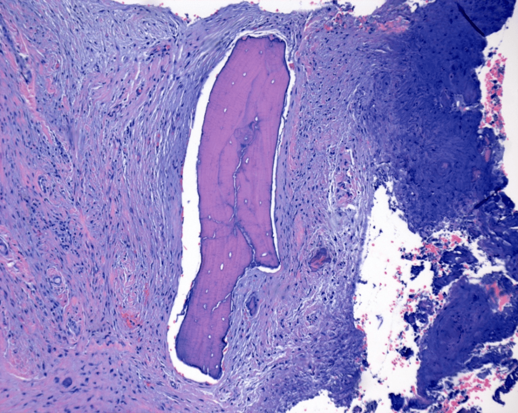
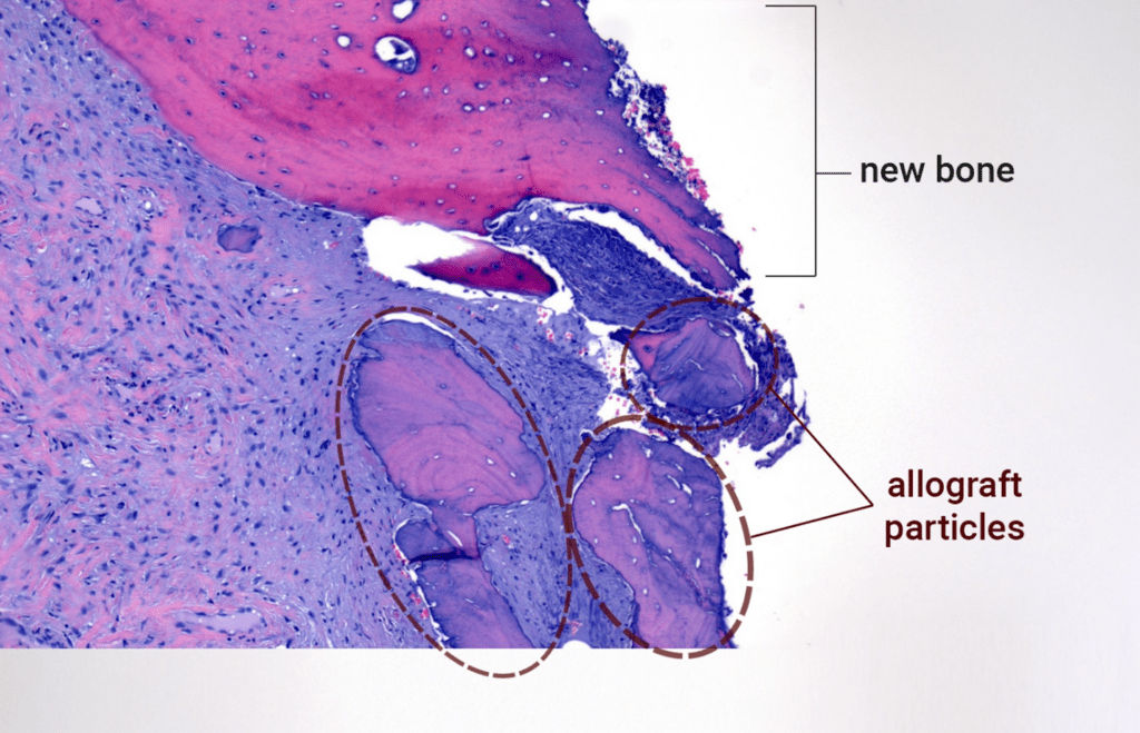
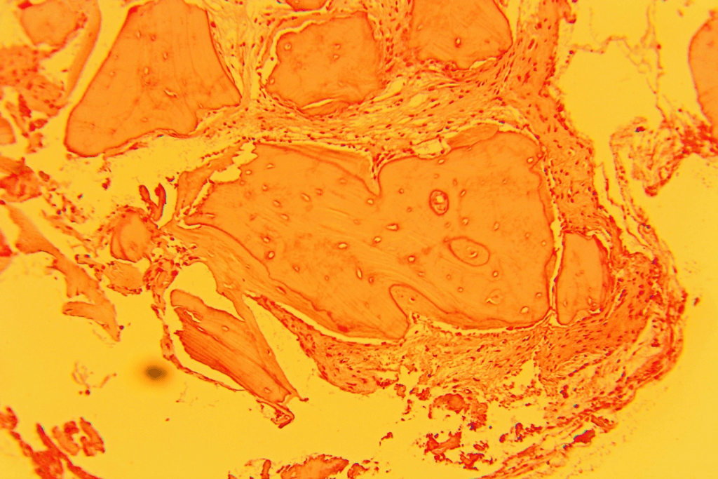
Distance intramembranous osteogenesis with osteoid-front mineralization combined with fibrous encapsulation of the allograft particles:

What these images show:

  • Allograft particles: large, acellular fragments with empty lacunae and sharp borders.
  • Interface: fibrous stroma is in direct apposition to the particle edges with some lining macrophages.
  • Surrounding tissue: collagen-rich connective tissue with spindle fibroblasts, scattered macrophages, a few small vessels; occasional erythrocytes.
  • Host vital bone plate (top): Continuous mineralized matrix with viable osteocytes in lacunae and occasional cement/reversal lines. No broad osteoid seams. This is mature compact bone, not an active forming front.

Interpretation: This socket area shows persistent MFDBA particles that are not being integrated or remodeled. They are separated by fibrous connective tissue from adjacent host bone, with low vascularity and no active apposition or resorption at the particle surface (i.e., a low-turnover, foreign-body–type interface with no bone formation on the graft).

Pericellular / Matrix-vesicle–Mediated Mineralization without Osteogenesis

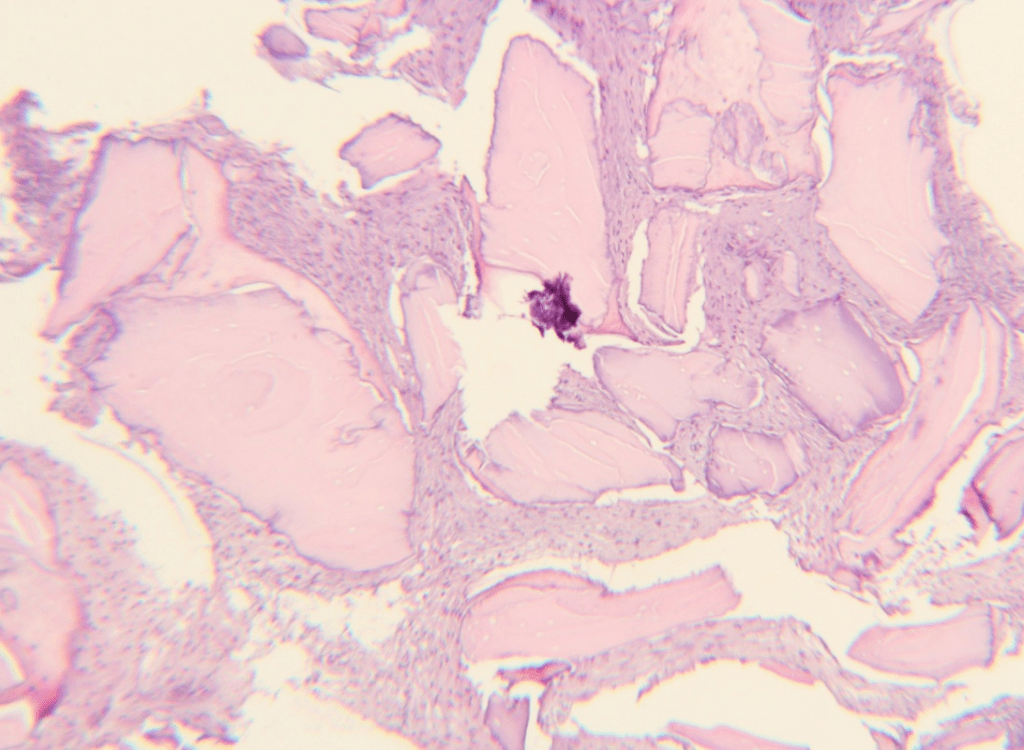
Bio-Oss one year after grafting:

Bio-Oss one year after grafting at low power.

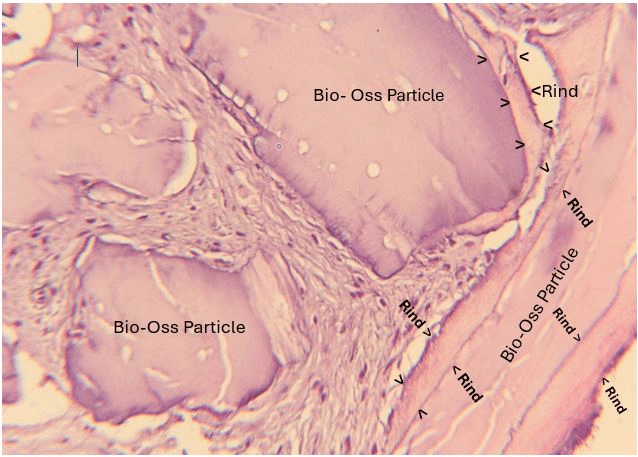
Bio-Oss one year after grafting at higher power

Interpretation: At one year, these fields are absent new bone formation on the Bio-Oss surfaces and no bone-to-implant contact. The graft particles appear persistent and largely un-remodeled, functioning as space maintainers rather than being replaced by vital bone.

The characterizing feature of this histology is the mineral rind that has formed on the Bio-Oss granules. The rind is produced by mineralizing macrophages that have been activated to mineralize the surface of the graft particles but this is not bone. The mineralization of the graft particles has stalled and is incomplete. Because the Bio-Oss particles are only partially covered with rind, no bone has formed in the socket after one year.

In the previous histology where no mineral deposit has occurred on the allograft particle, the particles remained isolated in fibrous tissue with normal bone growing around the allograft particles. In the Bio-Oss case, no bone formation has occurred with partial rind production. It appears from the histology that with minimal inflammation there is no reaction to the graft particles; however, without rind coverage of the particles bone formation never occurs in the socket. The cadaver bone graft particles only begin to be covered with bone after the rind has covered the particle. Once the rind covers the graft particle inflammation is reduced and osteoblasts can enter to begin to cover the rind with bone. This would be soft to drill into and implant integration would fail.

High power (interface detail)

  • The Bio-Oss surface shows smooth, acellular mineral with empty lacunae/micropores with rind formation.
  • The immediate interface is fibrovascular tissue with spindle fibroblasts and small vessels; no osteoid seam or lining osteoblasts abutting the particle
  • No Howship’s lacunae or osteoclasts → no active resorption of the granule

Interpretation: At one year, these fields are absent new bone formation on the Bio-Oss surfaces and no bone-to-implant contact. The graft particles appear persistent and largely un-remodeled, functioning as space maintainers rather than being replaced by vital bone.

The characterizing feature of this histology is the mineral rind that has formed on the Bio-Oss granules. The rind is produced by mineralizing macrophages that have been activated to mineralize the surface of the graft particles but this is not bone. The mineralization of the graft particles has stalled and is incomplete. Because the Bio-Oss particles are only partially covered with rind, no bone has formed in the socket after one year.

In the previous histology where no mineral deposit has occurred on the allograft particle, the particles remained isolated in fibrous tissue with normal bone growing around the allograft particles. In The Bio-Oss case, no bone formation has occurred with partial rind production. It appears from the histology that with minimal inflammation there is no reaction to the graft particles; however, without rind coverage of the particles bone formation never occurs in the socket. The cadaver bone graft particles only begin to be covered with bone after the rind has covered the particle. Once the rind covers the graft particle inflammation is reduced and ostoblasts can enter to begin to cover the rind with bone. This would be soft to drill into and implant integration would fail.

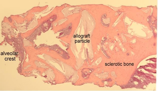
Allograft producing moderate inflammation.

Pericellular / matrix-vesicle–mediated mineralization of the allograft particles followed by distance intramembranous osteogenesis producing sclerotic bone without remodeling.

This is a high power of the mineralization process on an allograft particle.

Core (allograft particle):

  • Pale, strongly eosinophilic, acellular matrix with empty osteocyte lacunae.
  • Smooth, angular borders; no scalloped resorption bays, so no evidence of osteoclastic activity at this interface.

Immediate interface (rind):

  • A thin, hypermineralized rim hugging the particle.
  • This is an acellular mineral coat rather than true osteoid, consistent with dystrophic/mineralizing-macrophage mineral deposition that is not bone.

Cellular cuff around the rind:

  • Cells are foreign-body–type mineralizing macrophages
  • Where a lining cell sits, it’s discontinuous and not forming a uniform osteoid seam; no osteoblasts are found in this image.

Outer shell (host sclerotic compact bone):

  • Dense, bright eosinophilic sclerotic bone forming a circumferential encasement
  • Osteocytes are present but relatively sparse/irregularly spaced; canaliculi are not prominent
  • Surfaces are mostly smooth/quiescent; no true osteoid seams or active, continuous osteoblastic palisades
  • No osteoclasts identified on this field

Interpretation: a persistent MFDBA particle is wrapped by an acellular mineral rind and a macrophage-rich cellular cuff, all entombed in sclerotic compact bone with few osteocytes and no visible resorption. This supports a low-turnover, inflammation-associated mineralization that has been secondarily “overbuilt” by limited osteoblastic activity, rather than a healthy, vascular, remodeling bone front.

How this tissue forms:

  • Step A (weeks 1–4): Persistent particle triggers foreign-body inflammation → macrophages release TNAP and matrix vesicles → an acellular mineral rind precipitates directly on the graft surface(rind).
  • Step B (weeks 4–8): Inflammatory signals recruit/proximity-differentiate stromal cells; limited blood supply allows only a thin osteoid seam to be laid on the rind.
  • Step C (months 2–6): The interface remains cell-rich (macrophages + a few osteoblast-like cells), giving the dark purple band. With no osteoclastic turnover, the complex gets overbuilt and sclerotic rather than remodeled, ultimately entombing the particle.

This image shows an allograft particle 8 weeks after grafting. The particle is covered with a rind, and at this point, distance osteogenesis can begin with the formation of lamellar sclerotic bone, leading to the following image.

This is what this 6-month socket looks like histologically after grafted with mineralized freeze dried bone allograft:

  • Overall architecture: The space is largely filled by dense, sclerotic compact bone with very few marrow spaces. Trabeculae are thick and confluent, with multiple reversal/cement lines, indicating previous, patchy remodeling.
  • Allograft particles: Multiple pale, angular mineralized FDBA particles are still present. They are eosinophilic, often show empty osteocyte lacunae (non-vital matrix), and are encased by dense sclerotic host bone rather than fully replaced. Edges are sharp with little scalloping, consistent with limited resorption.
  • Host bone around the graft: Surrounding bone is predominantly lamellar/sclerotic with entombed osteocytes in lacunae; canaliculi are present but stroma spaces are markedly reduced.
  • Stroma/connective tissue spaces: Where spaces remain, they contain fibrous connective tissue rich in collagen with fibroblasts and sparse small vessels (endothelial cells). There is minimal granulation tissue.
  • Cells
    • Osteocytes within the sclerotic host bone (vital)
    • Empty lacunae within the allograft particles (non-vital)
    • No osteoblasts present
    • No osteoclasts present
    • Fibroblasts in intertrabecular fibrous tissue
    • Endothelial cells in a few small capillaries, low overall vascularity
    • Pericellular / matrix-vesicle–mediated mineralization via mineralizing macrophages
    • Overt inflammation is minimal

At 6 months, the socket is dominated by persistent MFDBA particles embedded in sclerotic compact bone, with limited stroma space, low vascularity, no osteoclast activity, and no osteoblasts. This image is consistent with an inflammation-driven, low-turnover mineralization rather than healthy osteoblast-mediated remodeling.
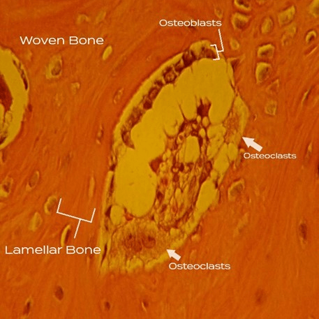
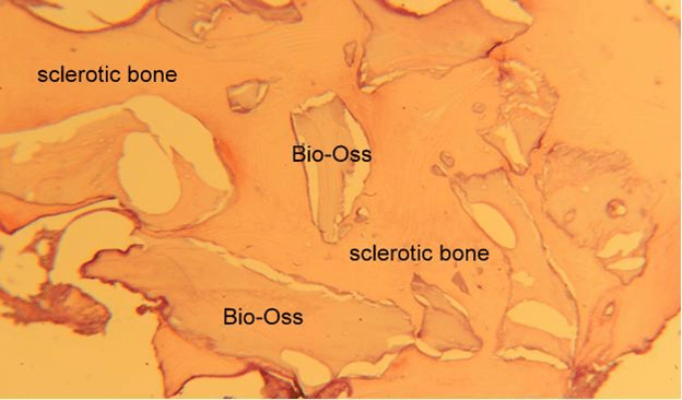
Just as moderate inflammation produced dense sclerotic with the allograft material, in this image Bio-Oss when producing moderate inflammation also produces dense sclerotic bone that never remodels.

Clinical Significance: Due to the inflammation produced by the transplanted graft material normal bone formation is blocked. In order to protect the host from the inflammatory material inflammatory macrophages that have been drawn to the area are recruited into mineralizing cells that encapsulate the offending graft material in a mineral rind. Once the rind is formed and the graft material is isolated, inflammation decreases and osteoprogenitors are now able to enter the area and produce bone. However, the rind does not produce the normal signals needed for the initiation of normal bone formation. Therefore, remodeling of the bone never occurs resulting in dense, brittle bone that cannot adapt to change and ultimately fails.

De Novo Intragraft Osteogenesis

Bone islands start inside the graft matrix. This type of osteogenesis occurs when bone producing cells crawl into a matrix and begin to randomly form bone in the matrix without being on a surface.

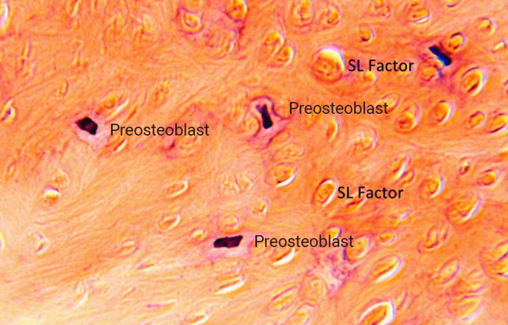
Socket grafted with Socket Graft Injectable after 2 days.

What the cells are doing: These are spindle-shaped osteoprogenitors moving through the porous spaces of the Socket Graft Injectable bone graft. The elongated nuclei and tapered cytoplasm suggest front-rear polarity. You can see fine processes extending toward and along graft surfaces, consistent with:

  • Adhesion and spreading on the material (integrin–mediated focal contacts)
  • Filopodia / lamellipodia probing pores to find paths of least resistance
    • Filopodia: Thin, spike-like membrane protrusions
    • Lamellipodia: Broad, sheet-like membrane ruffles at the leading edge
  • Osteoprogenator cells are fusing with vacuoles of SL Factor resulting is stimulating osteogenesis.

Socket grafted with Socket Graft Injectable after 4 weeks.

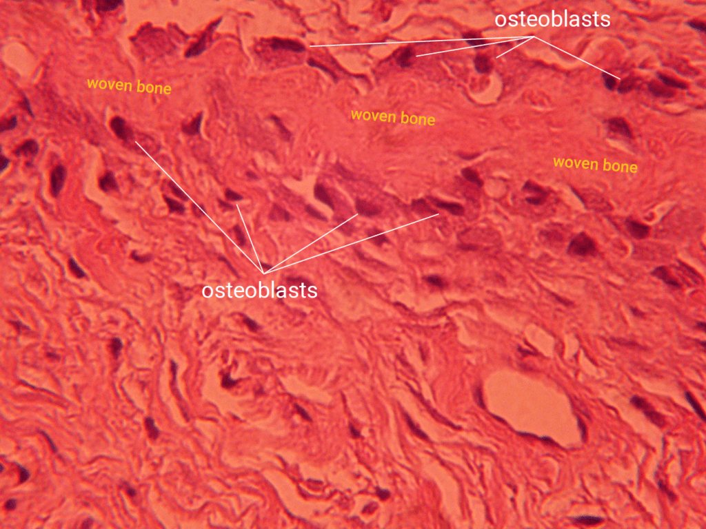
  • Dominant finding: Large, uniform eosinophilic osteoid masses within the SGI matrix. This is de novo osteoid filling intragranular spaces. The subtle parallel striations reflect appositional deposition by osteoblasts.
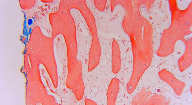
  • Rimming osteoblasts: Along the margins of these osteoid bodies are lined/clustered osteoblasts (oval to plump nuclei, polarized cytoplasm) consistent with active surface deposition. Several margins show softly scalloped growth fronts, typical of ongoing osteoid expansion.
  • Early entrapment: In a few areas there are faint lacunar silhouettes within the osteoid, suggesting early osteocyte entrapment (i.e., transition from osteoblast to osteocyte beginning inside the newly formed matrix).
  • Intervening stroma: Between osteoid-filled intragraft spaces is fibrovascular tissue (small vessels, stromal cells) that supplies and supports the osteogenic fronts.
  • Inflammation / foreign-body reaction: None evident in this field.

Interpretation: This field shows SL Factor-associated de novo osteoid formation by osteoblasts occurring inside SGI matrix (intragraft osteogenesis). The pattern is contact osteogenesis from within the graft rather than peripheral distance osteogenesis for the socket wall. Mineralization is just beginning/incipient here; more distinct mineralized fronts will appear in subsequent weeks as this osteoid matures into woven bone.

The same 4 week sample of socket grafted with Socket Graft Injectable consistent with de novo intragraft osteogenesis:

  • Tissue pattern: A broad sheet of immature osteoid/woven matrix with a wavy, fibrillar look. It’s punctuated by rounded/elliptic cell lacunae (early osteocytes) and small clusters of plump osteoblast-lineage cells at the upper margin.
  • Osteoid vs mineralized zones: Most of the field is light-to-mid eosinophilic osteoid; scattered denser, more glassy eosinophilic patches indicate incipient mineralization fronts within the osteoid. There isn’t a mature lamellar pattern yet.
  • Vessels / stroma: Interspersed fibrovascular channels feeding the osteogenic surface are present; no inflammation in this view.
  • Graft context: This osteoid occupies and bridges intra-particle spaces of the SGI (i.e., bone is forming inside the graft rather than creeping in only from the socket walls).

Mineralization process:

  1. Osteoblast secretion: Osteoblasts deposit type I–rich osteoid within SGI pores and along their internal surfaces, aligning with the wavy texture you see.
  2. Nucleation begins: Osteoblasts/early osteocytes release matrix vesicles and non-collagenous proteins (e.g., osteocalcin, osteopontin). In SGI + SL Factor, γ-carboxylation of osteocalcin supports Ca–P binding, favoring nucleation on/within the newly laid osteoid.
  3. Primary mineralization (ongoing here): Calcospherites (tiny mineral foci) form and coalesce, creating those denser eosinophilic islands/bands. Entrapped cells become early osteocytes.
  4. Secondary mineralization (to come): Over the next weeks-to-months, mineral content densifies and the tissue remodels from woven to lamellar bone and the Socket Graft Injectable graft material is resorbed.

At 4 weeks, this section shows active osteoblast-driven osteoid formation with the first islands/lines of mineral deposition inside the SGI, supported by good vascularity, and no foreign-body response.

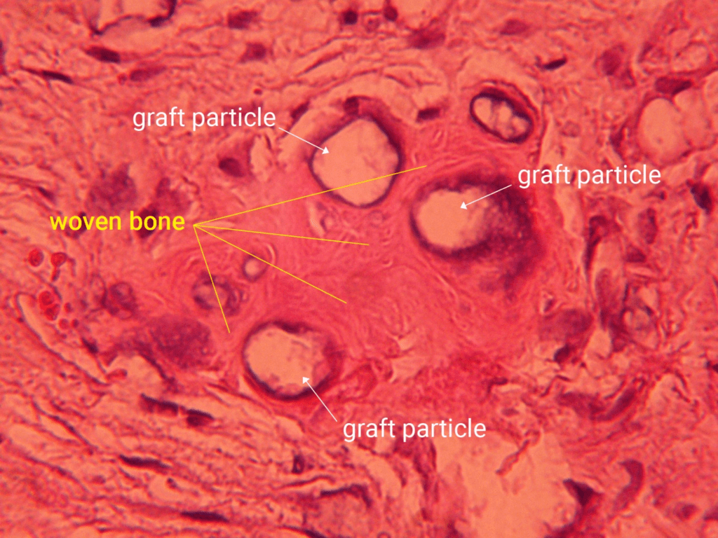
Histologic findings:

  • Former granule sites: Three central voids correspond to SGI CaP particles removed by demineralization during slide preparation.
  • Peri-void bone: Concentric rims of woven bone line each void; irregular, coarse matrix with variably spaced osteocyte lacunae. The rings represent successive osteoid deposition fronts, not true lamellae.
  • Interface line: A thin darker cement-line–like border at several void margins marks the prior mineral–tissue interface.
  • Matrix maturation: Surrounding areas show osteoid to early woven bone transitioning into denser but still non-lamellar bone; organized lamellar/compact architecture is not yet evident at this time point.
  • Background stroma: Fibrovascular connective tissue consistent with an actively healing socket.
  • Inflammation/FBGCs: Minimal to none; no conspicuous foreign-body giant cell reaction or fibrous encapsulation.

The pattern is most consistent with contact osteogenesis: osteoblasts have laid down concentric woven bone around the former SGI granules. At 4 weeks, this is expected; lamellar (compact) bone typically emerge later with remodeling. Overall, the section shows stable osteoconductive integration with ongoing maturation and no adverse inflammatory response.

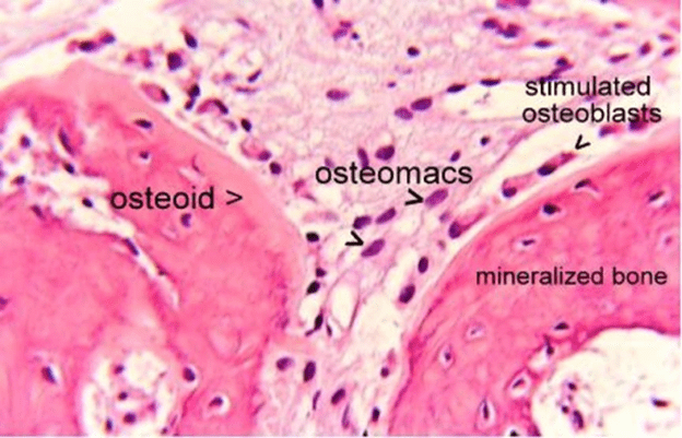
At 6 weeks this field shows an active osteoid-front mineralization during de novo intragraft osteogenesis:

  • Left: a broad osteoid seam (pale–eosinophilic, unmineralized matrix) with entrapped, newly embedded cells beginning the osteoblast→osteocyte transition.
  • Center interface: a narrow zone rich in osteomacs (osteal macrophages). They sit at the seam and “tend” the surface, clearing debris, supporting osteoblast survival, and coordinating mineral deposition at the front.
  • Right: mineralized woven bone with numerous osteocytes in lacunae, indicating prior osteoid that has already mineralized.
  • Lining cells: a row of stimulated osteoblasts rims the mineralized edge, showing polarized cytoplasm toward the matrix, consistent with continued appositional deposition.

Interpretation: Osteoblasts are laying down osteoid on the left; at the interface, osteomacs help maintain the osteogenic surface and couple it to mineralization; on the right, that osteoid has undergone primary mineralization (matrix‐vesicle–mediated nucleation, then calcospherite coalescence).

The overall direction of growth is from the mineralized side advancing into the osteoid, converting it to woven bone that will later remodel toward lamellar organization. Inflammation is absent, and the stroma above provides the vascular supply that supports these mineralizing osteoid fronts.

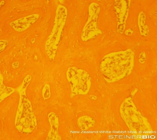
This image is 6 weeks after grafting with Socket Graft Injectable in the rabbit tibia. At this stage the image in filled with woven bone that is being remodeled into lamellar bone.

This high power image of the previous image shows a basic multicellular unit converting woven bone into lamellar bone. This is occurring because as bone forms in the tibia the graft becomes loaded as soon as the site fills with bone so the transition from woven bone to lamellar bone is rapid. This transition does not occur in the jaw until an implant is placed which then loads the bone and stimulates remodeling.

Six months after grafting a periodontal lesion with Socket Graft Injectable:

  • Periodontal side (left): A band of periodontal ligament (PDL) with dense, parallel collagen bundles. Fibers appear to insert into adjacent mineralized tissue as Sharpey’s fibers, consistent with a functional attachment.
  • Bone architecture: Predominantly mature lamellar bone forming interdigitating trabeculae. You can see:
    • Regular osteocyte lacunae within organized lamellae.
    • Occasional reversal/cement lines indicating prior remodeling fronts.
    • Stroma spaces between trabeculae filled with fibrovascular tissue (no inflammation).
  • Residual graft: In the lower-right lamellar bone there are small, discrete residual SGI particles. They are fully embedded in bone with contiguous matrix and nearby osteocyte lacunae; no fibrous encapsulation or foreign-body reaction.
  • Inflammation / giant cells: Absent; the stroma looks quiescent.

Interpretation: The site exhibits a mature, remodeled lamellar bone scaffold with healthy cellularity and active but orderly turnover (reversal lines). The PDL is preserved at the root surface, with fiber orientation consistent with normal function. Residual SGI is present as small islands but appears integrated within bone without adverse tissue response, which is compatible with gradual remodeling/resorption behavior over time. Overall, this image shows a stable, well-healed periodontal defect with restored hard-tissue architecture and a functional PDL The predictable way to maintain volume and produce healthy vital bone for long term implant success is with resorbable science-based synthetic bone graft materials.

SUMMARY

We have covered the main groups of bone grafts, how they produce bone and the type of bone they produce. The intent is for the reader to realize that the human body has miraculous methods of responding to different materials and how the body responds is predictable if you understand the material you are using. The only way to achieve a predictable result for your patients is to use a material that modulates its interaction with the host immune system and local regenerative cells to allow the normal mechanisms of bone formation and resorption to operate. The only type if bone that has been shown to provide long term support for dental implants in extraction sockets is healthy vital bone. The only way to maintain volume and predictably produce healthy vital bone is with resorbable science-based synthetic bone graft materials.

.

MEMBER:

American Society for Bone and Mineral Research (ASBMR)

Tissue Engineering and Regenerative Medicine International Society (TERMIS)

American Academy of Implant Dentistry (AAID)