Vertical Ridge Augmentation:

Failures and Breakthroughs

Decades of effort and surgical modifications have not improved the outcomes of lateral and vertical ridge augmentation. The failure of predictable ridge augmentation is not the failure of the surgeon. We have great surgeons. It is failure of the technology.

It is no surprise that the proposed unscrupulous scientific activities of a journal’s editor-in-chief would find its way into their journal. As you read this article by GenomeWeb, you’ll find that those who outed the editor made it clear that this editor was allowed to promote his fake science because dentists do not have a working knowledge of genetics, and that is precisely our point.

Here at SteinerBio predictable lateral ridge augmentation is routine:

The search for predictable lateral ridge augmentation is a thing of the past at SteinerBio. This post will share with you the exciting progress we are making with our goal of predictable vertical ridge augmentation.

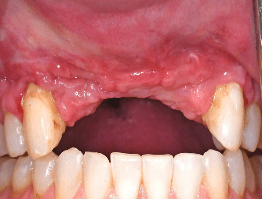
In the first case, significant bone loss occurred after a long standing implant had failed. Vertical ridge augmentation was required to restore function with new implants.

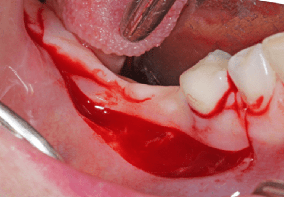
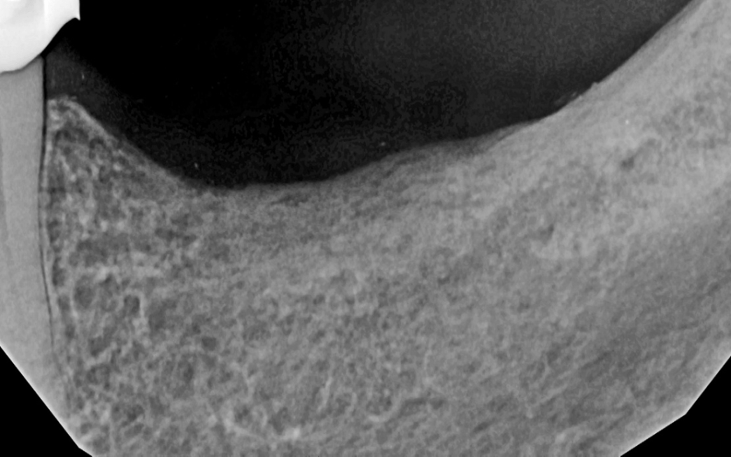
This is an area where a failed implant was removed.

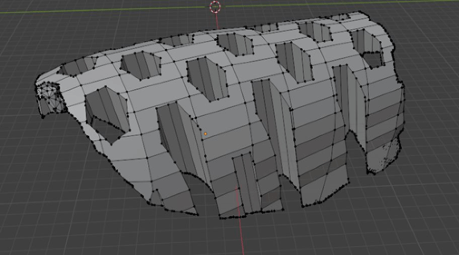
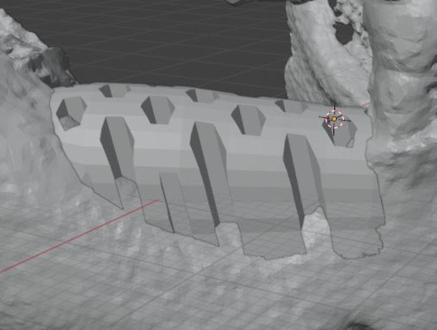
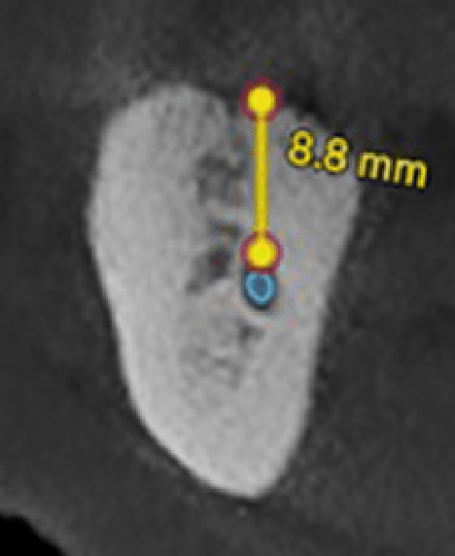
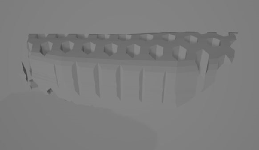
A ct scan showed 8 mm of bone coronal to the mandibular nerve. Vertical ridge augmentation was planned with a 3D printed graft.

Computer image of graft in place.

Traditionally, we use mucosal incisions and covering the graft with the periosteum for ridge augmentation (Simple Ridge Augmentation).

This is very successful in preventing graft exposure. However, the decision was made to use crestal incisions and use the inverted periosteal graft for covering the bone graft with the periosteum.

The periosteum is rich in bone regenerative cells and at SteinerBio, we have been using the inverted Periosteal Graft for the last 15 years to regenerate periodontal lesions. In this surgery, full thickness flaps are raised, the periosteum is dissected and sutured over the periodontal lesion with remarkable success. So, we thought this might be a method of covering the graft with the periosteum using a crestal incision.

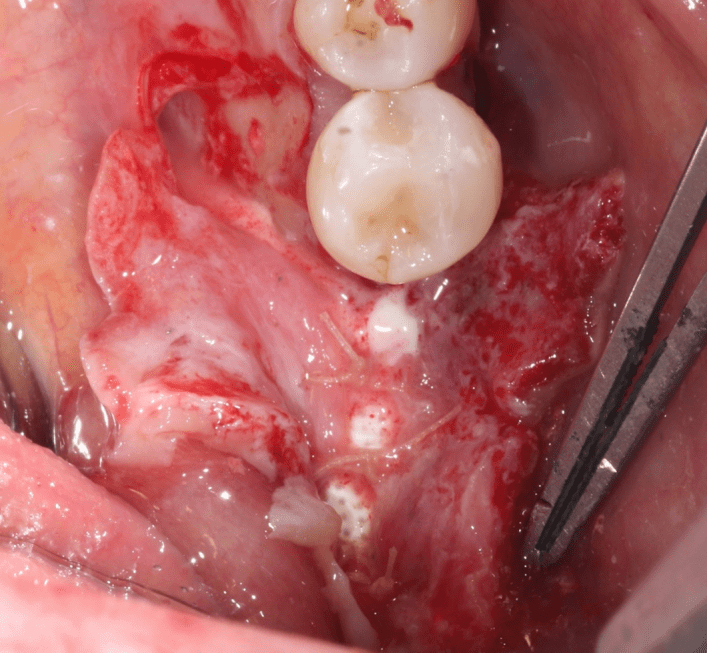
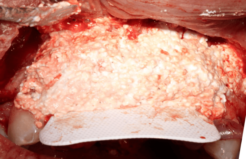
This image shows the 3D Printed Graft in place covered with a d-PTFE membrane and the periosteum being closed over the grafted site. After the periosteum is closed, the buccal and lingual flaps are sutured closed. When this procedure is done for treating periodontal disease just picture the areas of exposed membrane as the areas that would have teeth with the periosteum covering the graft material in the periodontal defect.

While this periodontal surgical procedure is far superior to anything being used today in the United States, the periosteum for regenerating periodontal lesions has received significant attention internationally. This procedure cannot be used with cadaver bone grafts and, therefore, periodontists in the US do not use this procedure.

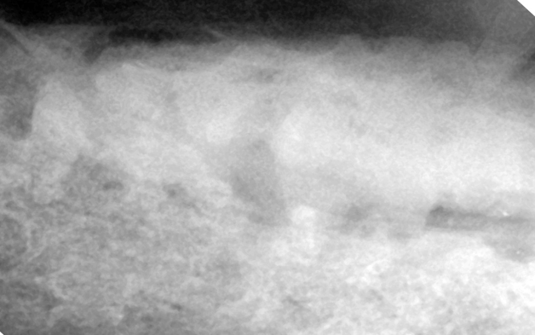
This image is two weeks post surgery. Over the alveolar bone is a dark radiolucent area which is showing active osteogenic invasion of the grafted site.

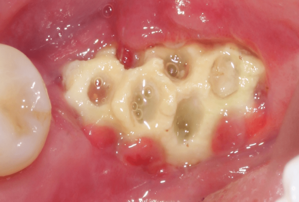
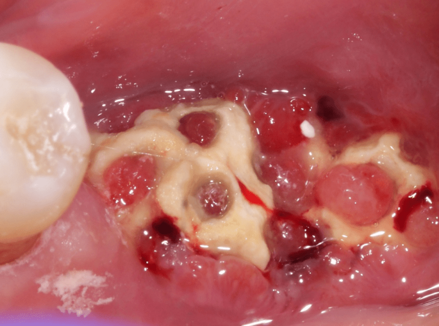
5 weeks post surgery shows exposure of the printed ridge with the assumption that the graft has failed. However, the graft is fixed and was obviously firmly connect to the underling bone. The decision was made to monitor the graft site. No antibiotics were prescribed.

At 2 months, the graft is solid and soft tissue is growing into the graft. Note the lack of bacterial accumulation.

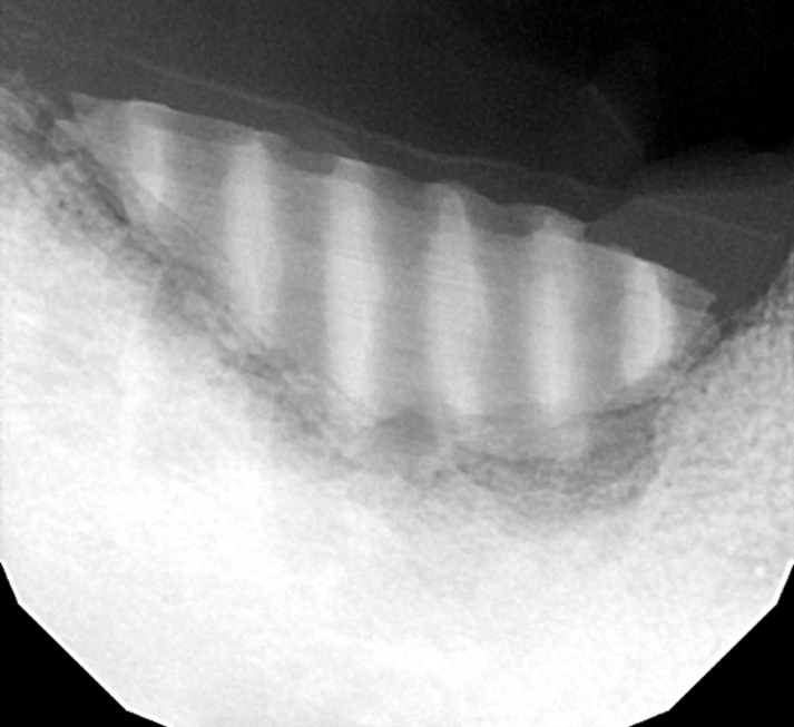
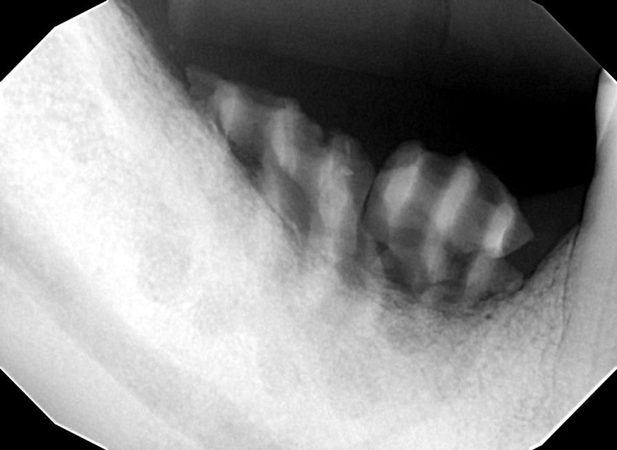
Radiograph at 2 months.

Radiograph at 3.5 months.

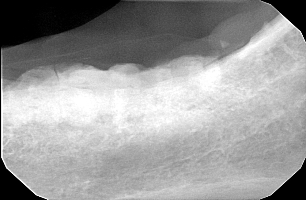
Radiograph at 4 months.

From failure to breakthrough:

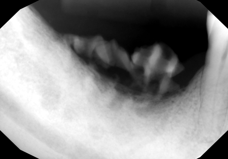
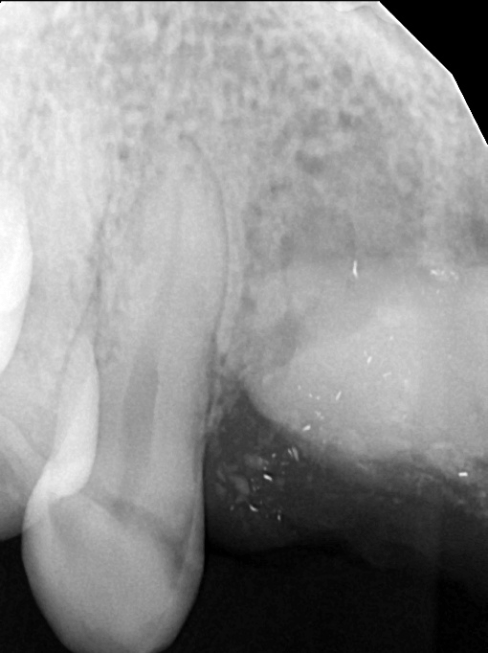
Anterior pre op

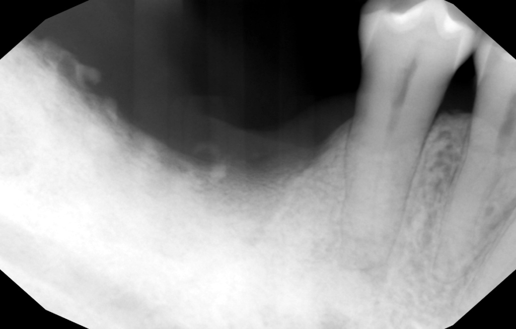
Anterior post op

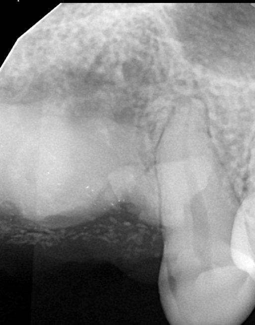
Posterior pre op

Posterior post op

So what happened?

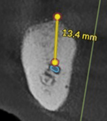
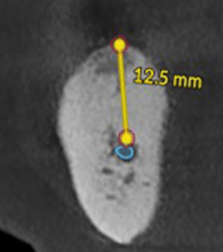
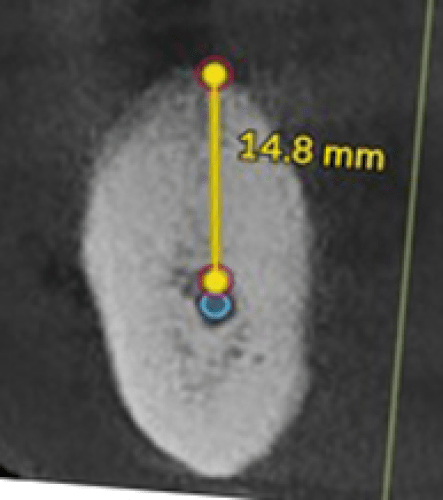
Clinically, the surgical design failed to cover the graft, but significant vertical bone regeneration was achieved. In the critical area, the bone above the nerve went from 8.8 mm to 13.4 mm, providing ample bone for full size implants.

How did the bone continue to grow into the graft and continue to regenerate bone when the graft was exposed to the oral cavity?

One factor is the design of the 3D print. It is composed of 100% pure beta TCP that is structured with interconnected micropores. These interconnected micropores are designed to be too small to be colonized, but because they are interconnected, they will produce a vigorous flow of extracellular fluid into and out of the graft. This explains why the graft did not become covered with plaque. It is also the reason why our beta TCP granules never get infected.

So what is the breakthrough?

The breakthrough is the vitality of the bone that was present when the graft was placed determined the ability of the graft to produce vertical ridge augmentation. The site was grafted with Socket Graft Plus when the implant was removed. Due to the stimulation of osteogenesis, this produces bone with a high concentration of regenerative cells. It was the realization that it is the vitality of the pre-existing bone that determines the success or failure of attempts to achieve vertical bone regeneration. The more vital the bone, the greater the potential for regeneration was the breakthrough. Please take a minute to look at the radiographs of the previous case and note the density of the bone.

After the breakthrough realization that you must have healthy vital bone for vertical ridge augmentation, we can now evaluate bone vitality radiographically and make a plan to prepare the site for vertical ridge augmentation.

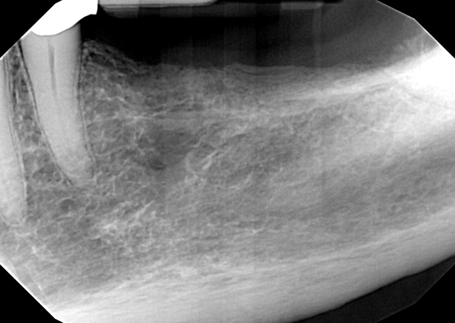
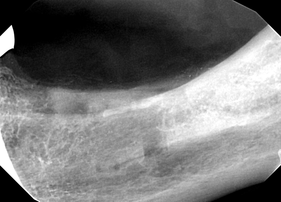
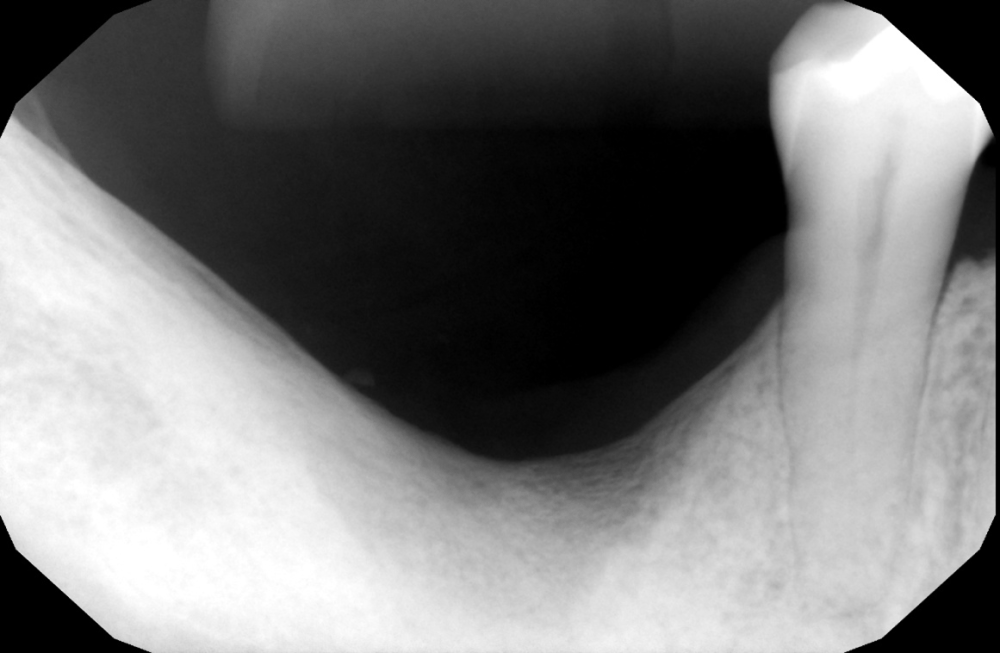
Preop radiograph. Years since the teeth have been extracted, the bone shows typical atrophy with a significant decrease in cancellous trabeculae. In addition, the porosity of the inferior border of the mandible is diagnostic for osteoporosis. While osteoporosis can lead to a reduction in mineral density, it has no effect on bone regeneration. Note the retained third molar.

In order to achieve vertical ridge augmentation, the bone must have a high level of vitality. In this case, the decision was made to remove the 3rd molar and regenerate the vitality of the bone. During the extraction, a crestal flap was raised and the crest of the bone was perforated the entire length of the edentulous area. The perforations into the cancellous bone was filled with Socket Graft Injectable. SteinerBio bone grafts are the only graft materials on the market with the ability to stimulate osteogenesis.

Day of surgery, after grafting the extraction socket and treating the edentulous bone with Socket Graft Injectable.

6 months after extraction of #17 and stimulating osteogenesis. Note the dense trabecular pattern and increase in radiopacity. The edentulous area now has a higher density and greater vitality that the preexisting bone distal to the bicuspid.

With bone vitality restored the 3D printed ridge is designed.

This radiograph is two weeks after grafting. Mucosal incisions were made and the periosteum was dissected. The bone was perforated and the site was covered with Socket Graft Injectable, which is used to bed the 3D printed graft. The graft is covered with Socket Graft Injectable and then covered with a d-PTFE membrane. The periosteum is sutured over the d-PTFE membrane, and then the mucosal incisions are closed.

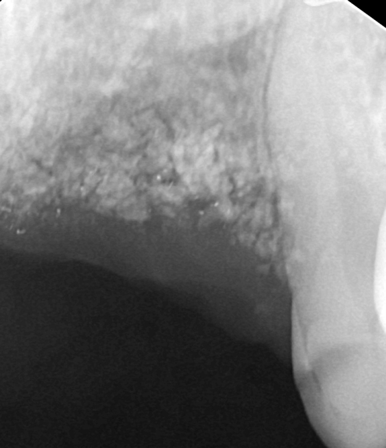
At two months, the graft shows surprising radiodensity throughout the graft.

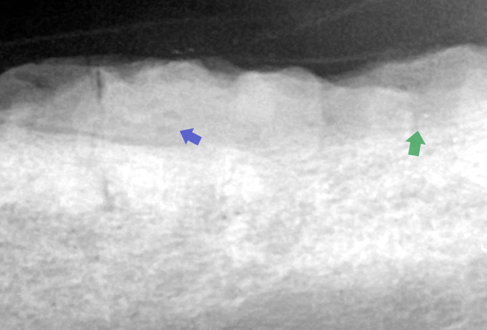
At 4 months, the graft is showing an increase in porosity (blue) and an increase in trabeculae formation (green) indicating remodeling of the 3D printed graft.

Each case presents with a different history and to determine the vitality of the bone what was done to the bone needs to be understood.

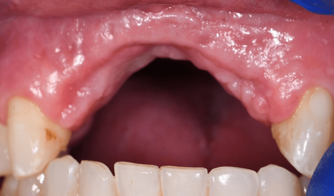
This case had an extensive history but most recently the area was grafted with Augma Bone graft material.

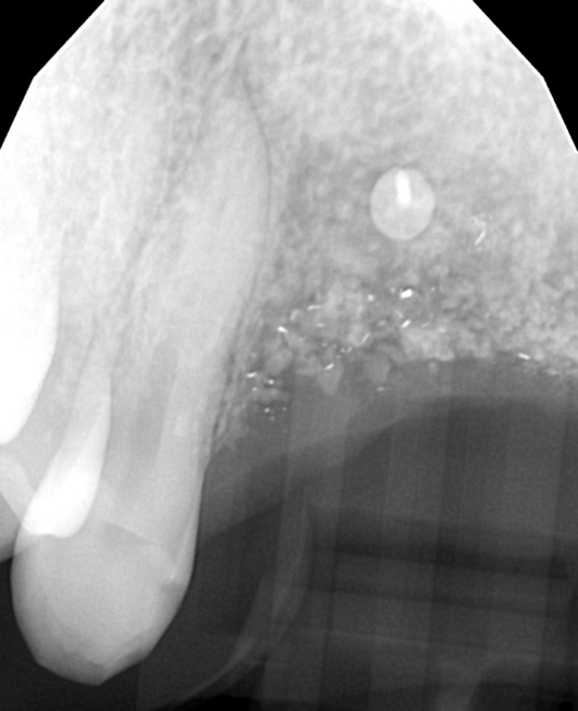
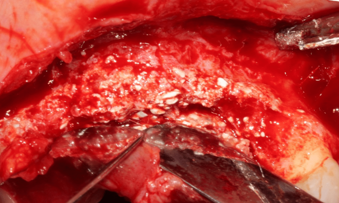
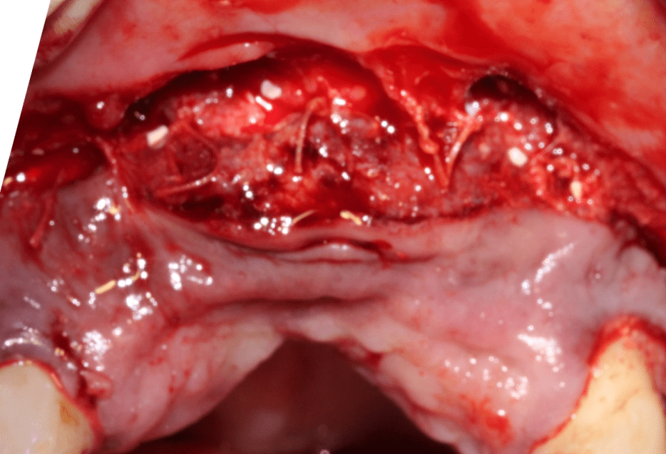
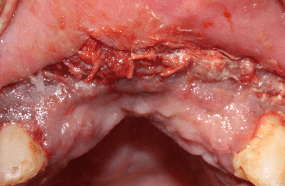
Augma Bone graft is nonresorbable and the graft site is filled with residual debris. This bone has not only suffered extensive trauma, but is also filled with debris from previous grafting, making it impossible to regenerate any new bone over what is severely compromised bone. In order to restore the vitality of the bone, the residual graft material needs to be removed and the underlying bone must be stimulated to restore vitality.

The surgical entry is the same as the previous case. After exposing the ridge, the residual Augma Bone graft is evident in fibrous connective tissue. The fibrous connective tissue over the ridge was removed with the removal of bone containing residual Augma Bone particles. The site was then grafted with Ridge Graft Kit, which is composed of a mixture of Socket Graft Injectable and OsseoConduct beta TCP granules. A d-PTFE membrane covers the graft.

The periosteum is sutured over the membrane.

The mucosa is closed over the periosteum and sealed with Oral Bond.

The goal of the first surgery is to revitalize the patient’s bone and produce bone that will contribute a high population of regenerative cells that will convert the 3D printed graft into vital bone.

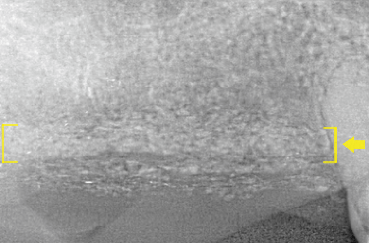
9 weeks after initial surgery. At this time frame, a new crest has formed (yellow) and the bone is regaining normal architecture. A significant amount of residual Augma Bone graft particles are present, but this graft material was embedded in the gingiva and was unable to be removed without compromising the vitality the gingival flaps. This material will eventually migrate out of the gingiva and into the mouth.

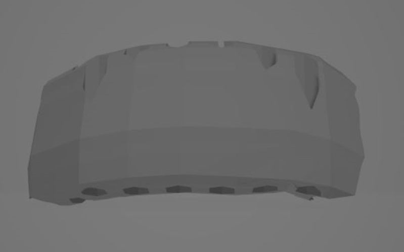
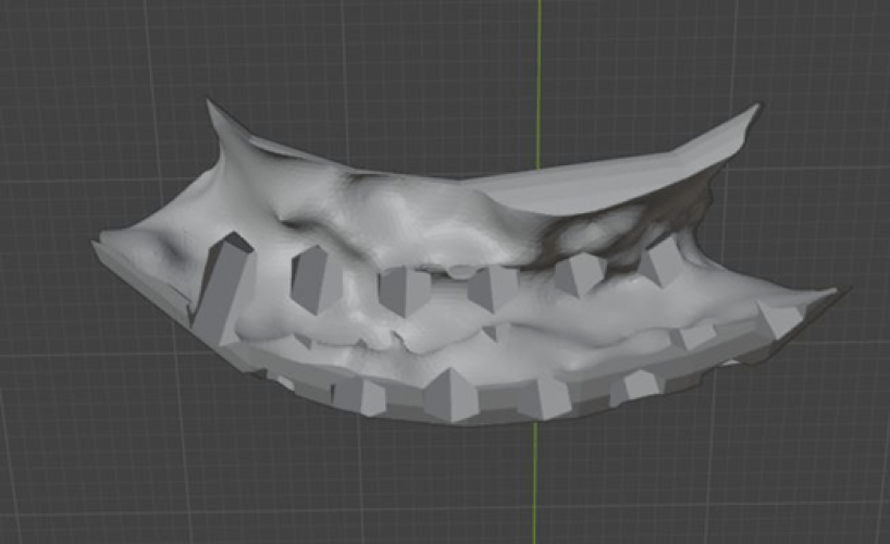
The 3D printed graft is designed.

Base of the graft.

Two weeks post op. Area of radiolucency between the bone and the graft shows aggressive proliferation of regenerative cells at this two week mark.

Two weeks post op. The surgical site remains closed as regeneration proceeds.

The points made in this discussion can only be used to guide you when using SteinerBio bone grafts because no other bone graft changes the physiology of the osteoblast and modifies vitality.

In addition, you cannot equate mineral density on a radiograph with an increase in vitality unless you understand the mechanism that produced the increase in radiopacity. SteinerBio bone grafts increase in radiopacity as mineralization proceeds, but it is also accompanied by a normal trabecular architecture and by the ability of the bone to change and remodel when placed under load.

Recent research shows even the most experienced and skilled surgeons fail at even lateral ridge augmentation when using traditional bone graft technology.
Volumetric assessment of changes in the alveolar ridge dimension following GBR using FDBA + collagen membrane or a novel resorbable collagen scaffold: a prospective two-center clinical trial.

  • J Periodontol. 2021;93(3):343–353. doi:10.1002/JPER.21-0006.
  • Dowlatshahi S, Chen C-Y, Zigdon-Giladi H, Horwitz J, Ahn C, Kim DM, Machtei EE

The cited paper is illustrative of the problem with using cadaver bone grafts for augmentation surgery. Allografts were used as a control, assuming that this material would produce a significant amount of lateral ridge augmentation. However, in this very well done study, lateral ridge augmentation produced on average less that 1 mm of bone augmentation. Our entire profession is taught that cadaver bone grafts are effective at lateral ridge augmentation but all to often these surgeries fail and an endless list of causes are listed except the cadaver graft material.

A well done German study found that failure of allografts is related to a systemic inflammatory response to the graft material.

Characterization of immunologically detectable T-cell sensitization, Immunohistochemical detection of pro-inflammatory cytokines, and clinical parameters of patients after allogeneic intraoral bone grafting procedures: a prospective randomized controlled clinical trial in humans

  • BMC Oral Health. 2022 Dec 10;22(1):592.
  • Solakoglu Ö, Götz W, von Baehr V, Heydecke G, Pantel K, Schwarzenbach H

In conclusion: when a systemic immune/inflammatory reaction occurred, the graft underperformed/failed and needed corrective surgery.

The German study concluded that unless you do tissue typing, grafting with allografts will never be predictable.

While cadaver bone grafts are not predictable, this presentation shows that synthetic bone grafts can also compromise bone and fail. The only tissue proven to support your implants with long term predictability is normal bone. If you graft with a nonresorable material such as the Augma Bone grafts used in the case above, you will never produce normal bone.

While a cadaver bone graft produces sclerosis via inflammation, nonresorbable bone grafts produce sclerosis due to the inability of the bone to remodel.

The success of vertical bone regeneration is not dependent on our 3D printed beta TCP bone graft. The critical factor is the mating of our 3D printed beta TCP bone grafts that are fully resorbable with our technology that stimulates osteogenesis.

The use of BioDensification for immediate implants produces integration in the grafted areas, avoiding angular defects and increasing implant success rates. Filling the osteotomy with BioDensification when poorly mineralized bone is encountered regenerates the surrounding bone and increases implant success rates. Many clinicians use this technique when using zirconia implants to achieve an integration rate equivalent to titanium implants.

Formulating a Predictable Protocol for Ridge Augmentation

  • Lateral ridge augmentation when only a few mm are needed for implant placement.
    • Use the Simple Ridge Augmentation procedure with SteinerBio Ridge Graft Kit and d-PTFE membrane
  • Significant lateral ridge augmentation cases
    • Use the 3D printed Osseoconduct Beta TCP ridge combined with Socket Graft Injectable
  • Vertical Ridge Augmentation
    • Prep the site with the Simple Ridge Augmentation procedure
    • Secondary surgery using the 3D printed Osseoconduct Beta TCP ridge combined with Socket Graft Injectable

For treatment planning with the 3D printed ridge click here:

MEMBER:

American Society for Bone and Mineral Research (ASBMR)

Tissue Engineering and Regenerative Medicine International Society (TERMIS)

American Academy of Implant Dentistry (AAID)